Introduction
The foot is where movement begins, from the initiating of simple functional movements such as sit to stand or walking, to climbing stairs, to more complex dynamic sport movements such as playing soccer, football, rugby, and tennis. The ankle and foot complex require proper mobility in order for the body to initiate movement or change direction. In this article, we will review the anatomy of the ankle, common injuries to the ankle, functional assessments and training strategies to work with clients with previous injuries.
Basic anatomy
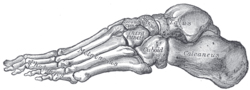
Fig 1. Metatarsals/phalanges(forefoot), Midfoot, Rear foot
Let’s look at the basic anatomy of the foot. There are three functional areas within the ankle; the forefoot(front), midfoot(form the arch) and the rear foot(back), which can be seen at right. The forefoot is composed of the five toes (called phalanges) and their connecting long bones (metatarsals). The midfoot has five irregularly shaped tarsal bones that forms the foot’s arch, serving as a shock absorber. The rear foot is comprised of top of the talus is connected to the two long bones of the lower leg (tibia and fibula), forming a hinge that allows the foot to move up and down. The heel bone (calcaneus) is the largest bone in the foot. It joins the talus to form the subtalar joint.
When we look at the support and stability within the ankle, the primary support comes from the ligaments within the ankle. There are several important ligaments that stabilize, and a few, particularly that are often injured more than others.
Primary ligament restraints of the talocrural joint
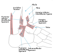
Fig 2. Ligaments of the foot
There are three primary ligaments that support the ankle, the anterior talofibular ligament, the calcaneofibular ligament and the deltoid ligament. These ligaments can be seen in figure 2.
a. Anterior talofibular(ATFL) ligament originates on front aspect of the fibula, 1 cm from the tip of the lateral malleolus and inserts along the outside aspect of the talar neck.
b. Calcaneofibular ligament(CFL) originates on the tip of the fibula, and inserts on the outside of the calcaneous(heel bone). It is the most often ligament injured due to being the weakness of ligament and because most ankle sprains involve inversion of the foot. (toe going inward). This is seen in figure 3.
c. Deltoid ligament originates from front & below aspects of medial malleolus and
inserts on medial and posteromedial aspect of the talus. It resists eversion motion of the foot.
Common injuries and causes
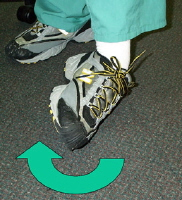
Fig 3. Inversion of the ankle
There are different types of injuries the ankle can sustain. The most common are the ankle sprain, plantar fasciitis and achilles tendonitis. In this next section, we will review each condition providing a deeper understanding of each.
a. Ankle Sprains
Mechanism of injury/pathophysiology: occurs as a result of direct trauma where 85-90% of all ankle sprains are lateral(inversion) ankle sprains, as seen in figure 3. With lateral sprains, the foot is plantar flexed and inverted at the time of injury injuring the anterior talofibular ligament (ATFL)initially, with the calcaneofibular ligament(CFL).
Sprains are graded from one to three based on the severity of the sprain and are as follows:
Grade 1: There is some loss of function with minimal tearing of the anterior talofibular ligament and mild swelling visibly present.
Grade 2: There is moderate loss of function, particularly with walking and negotiating uneven surfaces. Anatomically, there is partial tearing of the anterior talofibular ligament(ATFL) and calcaneofibular(CFL) ligaments, presenting moderate amount of swelling throughout the ankle.
Grade 3: There is severe loss of function, affecting a person’s ability to bear weight and walk due to severe pain and the complete tearing of the anterior talofibular ligament, calcaneofibular ligament and posterior talofibular ligaments. Clinically, there is a significant amount of swelling throughout the ankle.
Healing time of ligaments: Typically a grade one sprain, requires 0-4 weeks to completely heal, a grade 2, requires 4-8 weeks to heal
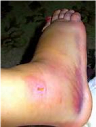
Fig. 4 Ankle sprain
and a grade three can take up to 12 weeks to heal and may require surgical intervention.
b. Plantar fascitis
Mechanism of injury/pathophysiology: Plantar fascitis is caused by repetitive mechanical loading of the plantar fascia, due to excessive pronation of the foot, resulting in an irritation and inflammation of the plantar fascia.
Risk Factors: Excessive pronation, decreased arch, unsupported shoes, muscle imbalance between the evertors and invertors.
Sign and symptoms: Pain occurs along the plantar fascia(typically along the medial aspect), with symptoms of gradual, insidious onset, with pain being worst in weight bearing, and localized heel pain as well.
Medical treatment: In the acute phase, physical therapy utilizes modalities to decrease inflammation and assist with tissue healing. In addition, taping, stretching, manual therapy and stabilization/strengthening exercises are utilized.
c. Achiles tendonitis
Mechanism of injury: The achilles tendon is among the most prone to overuse injury, with tendon problems accounting for up to 18% of injuries in runners (Magnussen et al 2009). Achilles tendonitis is defined as the acute inflammation of the tendon while an achilles tendinopathy, is defined as chronic pain in the achilles tendon. Injury to the Achilles tendon can be a result of overuse, commonly seen in sports that involve running and jumping. Excessive loading of the tendon during vigorous training activities is regarded as the main pathological stimulus or cause.
Achilles ruptures, typically affect younger individuals, in which running, jumping, and agility activities involving explosive, eccentric loading to the Achilles tendon. Natural aging allows predisposing chronic degeneration of the tendon. Blood flow decreases and stiffness increases with aging to decrease the ability to withstand stress(Hess, G 2010).
Pathophysiology: An overuse tendon injury, is caused by repetitive strain of the affected tendon such that the tendon can no longer endure tensile stress. As a result, tendon fibers begin to disrupt microscopically, leading to inflammation and pain(Paavola, M et al 2002).
Contributing factors: Training errors, running a distance that is to long, running too intense, increasing distance too greatly, excessive motion of the hind foot in the frontal plane, especially a lateral heel strike with excessive compensatory pronation, are all thought to cause or influence an achilles injury.
Sign and symptoms: Localized pain, the tendon is diffusely swollen and, on palpation, tenderness is usually greatest in its middle third. Chronic achilles injuries, will present with a tender, nodular swelling and pain again with dorsiflexion of the foot.
Medical treatment: Patients may be given initially NSAIDS for pain relief, are advised to rest, use ice and decrease the load to the foot complex. The role of corticosteroid injections in the treatment is controversial. Gentle stretching of gastrocnemius and conservative management is used for achilles tendonopathies. Diagnostically, ultrasound and MRI may be used to rule out tears.
Common assessments
The ankle is where all movement begins, therefore, possessing proper mobility is vital. Limitations in dorsiflexion can impair functional as well as sport movements. Several studies published have shown that limited dorsiflexion impacts the squat, step down(stairs), and landing from a jump. Therefore, lacking ankle mobility, particularly in the elderly predisposes them biomechanically to a fall.
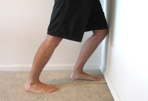
Fig 5. Standing wall test
One simple test to assess ankle mobility. It is the standing wall test, as seen in figure 5. In this test, have your client should be barefoot and begin in the standing position as if they were going to stretch their calf muscles. The lead foot should be 6” from the wall, this is important in standardizing the test. Measure and mark accordingly. From this position, have the client lean in, keeping their heel on the ground. From this position, you can measure the distance of the knee cap from the wall or measure the client’s great toe to the wall, watching for the heel to come off the floor.
Another great test to assess a client’s movement pattern, is the squat. The squat is a classic fundamental primal movement that someone typically performs almost on a daily basis. With this test, you can observe how the client’s ankle, knee, hip and back moves compared to normal movement patterns. This is seen in the figure below. If the client demonstrates excessive pronation of the foot, potential reason is due to weak evertors or is due to structural issue(pes plannus or flat feet). If the client demonstrates excessive supination of the foot, this could be due to tightness of peroneals and gastrocnemius, tightness of peroneals & iliotibilal band(ITB), structural changes of the foot(high arch).
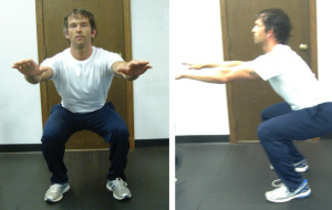
Fig 6. Squat in frontal view Squat in side view
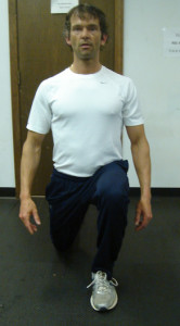
Fig 7. In place lunge
Lastly, an in place lunge looks at one’s control through the entire kinematic chain. The lunge is another fundamental primal movement. The lunge is a dynamic movement that is typically performed during daily activities(stooping down to pick something up) or as part of an athletic movement. This test examines ankle control, knee control and pelvic stability.
Training strategies and programming for ankle injuries
With any injury, the most important thing to remember is the type of injury, healing time and prior level of function of the client. Let’s begin with ankle sprains.
a. Ankle sprains
Recommendations for training: A client with a chronic ankle sprain would benefit from continued functional strengthening exercises that target the invertors, evertors and dorsiflexors. Exercises such as diagonal forward, diagonal reverse(as seen in figure 8, target the glute medius and minimus). In addition forward traveling lunges are effective in strengthening not only the ankle, but the knee and hip complex.
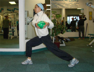
Fig 8. Diagonal lunge
b. Plantar fascitis
Recommendations for training: Strengthening the intrinsic and extrinsic muscles (evertors and invertors), is important. It is also important to continue stretching tight hip flexors, quadriceps, ITB and gastrocnemius. Core strengthening should always be part of the training program, begin teaching static exercises, such as planks, trunk rotation with tubing, then progress to dynamic exercises such as traveling lunges with trunk rotation and diagonal lunge with wood chop.
c. Achilles tendonitis
Recommendations for training: It has been shown through the research, that
12 weeks of eccentric resistance training can reduce pain in those suffering from
chronic achilles tendinosis. Physiologically, eccentric training stimulates type I collagen synthesis, whereby strengthening the tendon, resulting in also reduction in pain in the tendon during loading.(Langberg, H et al 2007). An effective eccentric exercise for the achilles, is to place a strong resistance based band under the client’s ball of their foot, and step down(dorsiflexion of foot) off a 6-8” step, or something that is comfortable. Continued strengthening the entire kinematic chain is effective as well as recommending to the client to cross train with such interventions such as yoga, pilates, swimming, cycling and hiking.
Summary
The ankle is a complex unit that is comprised of a multitude of ligaments, tendons,
connective tissue, muscles that synergistically initiate and correct movement, and stabilize when an unstable environment. Understanding the anatomy, biomechanics and weak links of the ankle, common injuries and evidenced based training strategies, should provide you with the insight to better understand and work with clients with these kind of injuries more confidently.
Written by Chris Gellert, PT, MMusc & Sportsphysio, MPT, CSCS, AMS.
Chris is the CEO of Pinnacle Training & Consulting Systems(PTCS). A continuing
education company, that provides educational material in the forms of home study courses, live seminars, DVDs, webinars, articles and min books teaching in-depth, the foundation science, functional assessments and practical application behind Human Movement, that is evidenced based. Chris is both a dynamic physical therapist with 14 years experience, and a personal trainer with 17 years experience, with advanced training, has created over 10 courses, is an experienced international fitness presenter, writes for various websites and international publications, consults and teaches seminars on human movement. For more information, please visit www.pinnacle-tcs.com
REFERENCES
Hess, G., 2010,‘Achilles Tendon Rupture: A Review of Etiology, Population, Anatomy, Risk Factors, and Injury Prevention,’ Foot Ankle Specialist, vol. 3, no.1, pp. 29-32.
Langberg, H., et al 2007,’ ‘Eccentric rehabilitation exercise increases peritendinous type I collagen synthesis in humans with Achilles tendinosis,’ Scandavian Journal Medicine Science Sports, vo1. 17, pp. 61–66
Magnussen, R., et al., 2009, ‘Nonoperative Treatment of Midportion Achilles Tendinopathy: A Systematic Review,’ Clinical Journal Sports Medicine, vol. 19, number 1, pp. 54-63.
Mcpoil, T., et al 2008,’ Heel Pain—Plantar Fasciitis: Clinic Practice Guidelines Linked to the International Classification of Functioning, Disability, and Health from the American Physical Therapy Association, ‘ JOSPT, vol. 38, issue, 4, pp. 2-17.
Paavola, M., et al 2002,’ Current Concepts Review: Achilles Tendinopathy,’ The Journal of Bone and Joint Surgery, pp. 2062-2073.
Wearing et al., 2006, ‘The pathomechanics of plantar fasciitis,’ Sports Medicine, vol. 36, issue 7, pp. 585–611.