Hide

Error message here!

Lost your password? Please enter your email address. You will receive a link to create a new password.

Error message here!

Back to log-in

Close
aging-hands

The “Dark Side” of Parkinson’s Disease: Why Parkinson’s Changed The Way We See, Part 1

The dark side of the moon is not considered a particularly exciting topic unless you are an astronomer or a Pink Floyd fan.  However, the dark side of the moon exists and is just as important as the front of it.

So, what are the conditions of the dark side of the moon? Does it ever see the light of the sun?  Why is it called the dark side?  These are interesting questions with answers that I will leave to space experts, BUT what I will say is that the moon is similar to Parkinson’s Disease (PD).  Yes, you read that correctly — the moon and Parkinson’s Disease are similar.

Parkinson’s Disease is most often characterized by a tremor and/or gait issues.  Just as the moon has a dark side, unseen but present, Parkinson’s Disease has a dark side”.  As a fitness professional, understanding the “dark side” of Parkinson’s Disease and how it affects your “fighters” will enhance your program design skills, increase your confidence when counseling a “fighter” or care-partner and help build your relationship with other medical and fitness professionals.

The goal of this article series is to shed light on the “dark side” of PD. By incorporating the knowledge of medical professionals on my Bridges For Parkinson’s team, you will learn about research discoveries, resources for program design, useful tools for “fighters” and care-partners and ultimately, hope! 

Vision impairments in those with PD is our first topic in this series. People living with Parkinson’s Disease experience changes in their vision as they age. These changes may include cataracts, macular degeneration, dry eyes, floaters, glaucoma, diabetic retinopathy, detached retina, trichiasis, and blepharitis. NOW, People living with PD may experience one or several of these issues due to normal aging. However, due to the depletion of dopamine neurons in the substantia nigra, those with PD may also experience the following vision issues and/or impairments.

  1. Blepharospasm: uncontrollable eye twitching
  2. Blepharitis: Lack of blinking. On average, we blink 12-14x per minute. A person with PD may only blink 3-5x per minute. The result is inflammation on the edge of the eyelid. Eyelids may become irritated or itchy and appear greasy or crusty.
  3. Apraxia: “oculomotor apraxia” –  the absence of or defect in voluntary eye movement. This may result in trouble initiating movement and moving the eyes in a desired direction
  4. Diplopia (Double Vision): when a single object becomes two objects.
  5. Dry Eyes: eyes may feel sandy or gritty.
  6. Blurry Vision: MAY be related to dopamine depletion in back of the eye and within  the visual connections throughout the eye (Davis Phinney)
  7. Eye movement issues:
  8. Pursuit Movement: eyes are unable to work together to follow an object such as a plane flying across the sky.
  9. Saccadic Movement: unable to move eyes rapidly from one object to another such as completing a line in a book and going to the other side to the beginning of  the next line.
  10. Vergence Movement: as the target or object draws closer to the person with PD, the eyes are unable to converge to maintain focus on the object causing double vision. Approximately 30% of people living with PD suffer from Vergence Eye Movement problems.
  11. Depth/Distance Perception
  12. Photophobia (also known as Light Sensitivity): may wear sunglasses indoors due to bright lights.

Fitness Professionals should inquire about vision impairments before a “fighter” begins an exercise program and obtain specific information about the vision impairment. Each symptom listed above can potentially compromise gait/balance, increase difficulty with Activities of Daily Living (ADL) and cause those with PD to isolate or become depressed due to lack of independence. 

How We See 

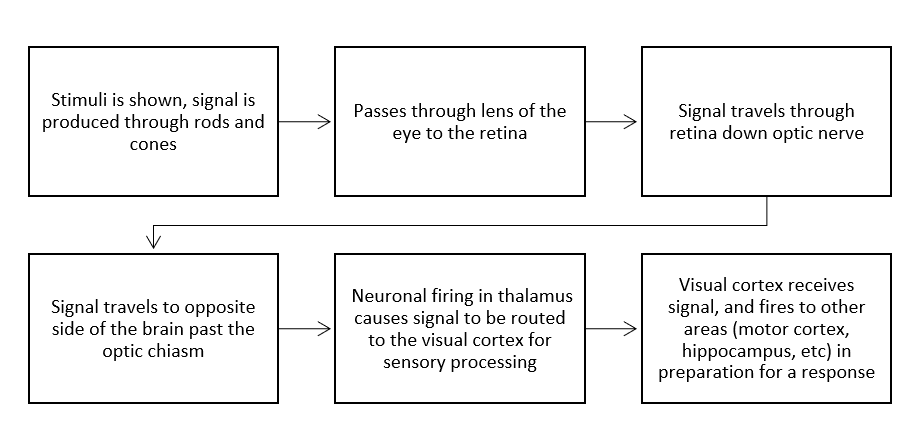
When we use our vision, we’re doing a lot of simultaneous tasks. We have to sense and process the stimuli, perceive and make sense of the stimuli in the context of the situation, and translate those signals into a response (i.e. movement, bringing up memories, emotions) to the initial stimuli. 

For example, a stimulus such as a car passing by causes numerous neurological responses to take place in the visual system. First, that stimulus passes through the lens into rod and cone cells that make up light/shadows and colors to put the image together, which then reaches axons in the retina that extend through the optic nerve. Once that signal travels through the optic nerve, it reaches the optic chiasm, where the stimulus passes over to the opposite side of the brain from the visual field and eye. Interestingly, this car is not visually processed right-side up! Instead, it goes into your brain upside-down as the eye lens flips the image. That image then travels through the thalamus (our “sensory relay system”), and to the primary visual cortex, where the image gets processed and relayed to other sensory systems to prepare for a response.The Role of Dopamine in the Eye
You wouldn’t think that dopamine is involved in vision. We think of it relative to its impact on motor control and behavior reinforcement. However, even in ocular movement, lack of dopamine can impact a person with PD significantly, possibly resulting in apraxia. Dopamine also regulates light sensitivity in our retinas that helps to modulate our circadian rhythm, known as our “internal clock”. As dopamine decreases with disease progression, the sensitivity of the visual system decreases as well, hindering the ability to differentiate between light conditions such as day and night, potentially leading to other problems in circadian rhythm (Witkovsky, 2003). Over time, we see a loss of dopamine projections into the eye worsening vision such as blurry vision, light sensitivity, or even color vision deficiency as listed above. Unfortunately, there is not enough research on the involvement of dopamine in PD vision to give a clear-cut role of dopamine, but current research points to dopamine as a larger part of vision problems experienced by those with PD.  

Physicians

It is crucial to understand the connection between the brain and PD vision related issues.  Knowing when to refer a “fighter” to a vision specialist is critical to building your team of advisors and establishing a strong medical-fitness program in your community.

I highly recommend finding a neuro-ophthalmologist or a neuro-rehab optometrist to join your team. A neuro-ophthalmologist specializes in both fields of neurology and ophthalmology.  They complete a residency in either neurology or ophthalmology then continue to complete a fellowship in the complementary field.

Dr. Jamie Ho, OD, FAAO, FCOVD of Nashville, TN is a member of my advisory board and she is a neuro-rehab optometrist. Dr. Ho addresses the functional deficits that result from the neurologic changes.  You can learn more about Dr Ho’s practice at www.hovisiongroup.com.

To find a neuro-optometrist or neuro-ophthalmologist in your area go to www.noravisionrehab.org 

Medication

Medication for PD certainly improves PD symptoms however, visual side-effects can occur.

For example, according to www.healio.com, Anticholinergic meds such as Artane are used to address tremors but can also cause dry or blurry vision. The Journal of Parkinson’s Disease notes that dopamine agonists cause hallucinations, Levodopa may lead to ocular dyskinesia (involuntary eye movements), and MAO inhibitors to blurry vision. 

Fitness Professionals need to record all medications during the initial assessment and update any medication changes quarterly, at minimum

Click to read Part Two of this article, which offers some tips for fitness professionals working with PD clients with vision challenges.


Co-authored by Colleen Bridges, M. Ed, NSCA-CPT; Renee Rouleau-B.S., PhD student, Jacobs School of Biomedical Sciences, University at Buffalo; Kristi Ramsey, OTD, OTR/L.

Reviewed By: Dr. Jamie Ho, OD, FAAO, FCOVD


References

  1. Berliner JM, Kluger BM, Corcos DM, Pelak VS, Gisbert R, McRae C, Atkinson CC, Schenkman M (2018) Patient perceptions of visual, vestibular, and oculomotor deficits in people with Parkinson’s disease. Physiother Theory Pract. doi: 10.1080/09593985.2018.1492055 [PubMed].
  2. Borm, C., Smilowska, K., de Vries, N. M., Bloem, B. R., & Theelen, T. (2019). How I do it: The Neuro-Ophthalmological Assessment in Parkinson’s Disease. Journal of Parkinson’s disease, 9(2), 427–435. https://doi.org/10.3233/JPD-181523
  3. www.DavidPhinneyFoundation.org.
  4. Mayo Clinic, (2020)“Parkinson’s Disease Symptoms and Causes.” http://wwwmayoclinic.org20376055. 
  5. Nowacka B, Lubinski W, Honczarenko K, Potemkowski A, Safranow K (2014) Ophthalmological features of Parkinson disease. Med Sci Monit 20, 2243–2249.
  6. Witkovsky, P. Dopamine and retinal function. Doc Ophthalmol 108, 17–39 (2004). https://doi.org/10.1023/B:DOOP.0000019487.88486.0a.
PDFS-Exercise

Bilateral Coordination: The Gateway to Successful Movement | Part 2

In Part 1 of this series, we discussed an overview of bilateral coordination, its importance and how it falters in Parkinson’s Disease (PD).

Now, we’ll discuss strengthening the pathways for those with PD with exercise.

Steps To Incorporating Bilateral Coordination Into Your Exercise Program

Clearly, the brain is a work of art when you consider the “architecture”, the “highway” of nerves required to communicate with the rest of the body, to the final outcome of the original thought or idea. ANY kind of “road block” is going to hinder an individual from completing tasks as simple as writing or buttoning a shirt. And for the person living with PD, this includes walking, bathing, driving, communicating, dressing, well, about every Activity of Daily Living (ADL) that you can conceive.

BUT… never fear, the PD Fitness Specialist is prepared to address these matters of the brain with some challenging YET fun activities to promote improved motor control!

I won’t lie, you may see smoke coming out of the “fighters” ears but the incredible sense of accomplishment at the completion of the drill will be worth it.

Always begin with the fundamental question of program design. What are the needs of my private clients and fighters? What are their common issues?  They definitely need to work on:

Strength (7 foundational movements)

  • Lunge
  • Squat
  • Pull
  • Push
  • Carry
  • Hinge
  • Rotate

NOTE: I encourage Fitness Professionals (FP) to start with the most basic form of each Foundational Movement before progressing to a more challenging version. I have learned that repetition and exercise phases are a necessary part of any fitness program, similar to the human development process.

Your program should also include cardiovascular endurance, agility (footwork/hand-eye), cognitive challenges, fine motor drills, balance/gait drills — ALL which incorporate Bilateral Coordination challenges that provoke the brain to enhance:

  1. Neuro-protection to preserve at-risk dopamine neurons.
  2. Neuro-repair to improve damaged “circuitry”  and rewire the brain.
  3. Neuro-Adaptation that trains the brain to move without conscious awareness of each move such as walking .

Yes, this requires the Fitness Professional to sit down and develop a program that is constantly evolving as the abilities and needs of the client change BUT it can be done. For example, to address gait and incorporate an additional Bilateral Coordination drill that will challenge your client(s) mental focus, try the following progressive drill.

NOTE: Step one is a fantastic way to help a person living with Parkinson’s Disease safely transition out of a “freezing of gait” moment.

Criss-Cross Applesauce (Stand in squat stance)-

  1. Cross the right hand to the left shoulder.
  2. Cross the left hand to the right shoulder.
  3. NOW, cross the right hand to the left knee.
  4. Cross the left hand to the right knee

Have your client(s) say “Criss-Cross Applesauce” while performing the drill. This will address hypophonia problems and assist in maintaining a strong beat..

Once they have achieved this version, have them progress to the next level….

Criss-Cross Applesauce with Marching Knees

  1. Cross the right hand to the left shoulder.
  2. Cross the left hand to the right shoulder.
  3. NOW, cross the right hand to the left knee BUT lift the knee to meet the hand as if marching.
  4. Cross the left hand to the right knee and lift knee to meet hand as if marching.

To add complexity to the drill, have your clients tap the marching knee onto a step or bosu.

Once they have achieved this version, have them progress to the next level….

Criss-Cross Applesauce with A Forward Lunge

  1. Cross the right hand to the left shoulder.
  2. Cross the left hand to the right shoulder
  3. NOW, cross the right hand to the left leg AS you lunge forward.
  4. Come back to start position.
  5. Cross left hand to the right leg AS you lunge forward
  6. Return to start and repeat drill

To add complexity to the drill, have your clients perform a diagonal lunge or a lateral step.

This is just one example of how you can incorporate Bilateral Coordination into a movement we do all day every day! Walking! And if you work with people living with Parkinson’s Disease, then expect their learning time to vary but with repetition and encouragement, they will conquer this drill and be excited to try the next.

Which leads me to share with you the results I have experienced in my Parkinson’s Disease Wellness Center in Nashville and Franklin, Tennessee.

CASE STUDY – RESULTS!

“Susan” is 62 years old and was diagnosed 17 years ago which classifies her as Young Onset Parkinson’s Disease.  Susan had the DBS surgery 10 years ago and although the DBS initially provided relief of tremors and dyskinesia, over time fine motor skills, drooling, hypophonia, balance/poor posture — leading to numerous falls — has become an issue. She is also blind in one eye which limits her spatial awareness, decreases balance and mobility, all of which makes living independently even more challenging. Additionally, the hypophonia had led her to become 80% non-verbal. To answer questions she either nodded yes/no or shrugged her shoulders if she didn’t know the answer.

When we began working 1:1 together, my first priority was to address her posture/gait, as she was stooped forward and shuffling, leading to multiple falls each week. So, in addition to a dynamic warm-up with large ROM drills to properly prepare her body from head to toe, strength training, boxing and cycling, obstacle courses and more, I taught her the “Criss-Cross Applesauce” drill. The first several sessions, Susan had to complete the drill 5x between other warm-up exercises AND march while tapping her hand to the opposite knee when we moved to a new station or machine. She also had to speak the words “Criss-Cross Applesauce” when performing the drill to address her hypophonia.

The first session, Susan could not make the connection that her hand was to tap the opposite shoulder or knee. I had to manually move her hands and say the words with her. By the end of the first session, she was only able to complete the drill at a slow tempo, but that was ok, she did it! She left the gym that day with homework to practice the “Criss-Cross Applesauce” drill three times a day for 5 repetitions. I also assigned marching in place while tapping the hand to the opposite knee 60x twice a day.

The second session, I noticed a significant difference in her timing and coordination. For the first set, I still needed to “mirror” her while she did the “Criss-Cross Applesauce” drill, but overall, Susan was able to complete the drill 3 out of 5x correctly. When Susan would move to a different location, I had her march and tap her hand to the opposite knee. We counted how many steps it took to make it to the next station with the goal of trimming 10-15 steps off the next round. To do so, I had her focus on making precise connections between her hand and opposite knee as well as stomping her foot when stepping. By the end of the session she was able to trim 5-10 steps off between stations.  She completed the session with the same homework as before.

The third session is when I started to notice some fantastic improvements. Susan walked into the gym marching and tapping the hand to the opposite knee. She was able to cover more ground with fewer steps and the best part was that stomping her foot was helping her step with increased assurance. That equates to fewer falls! Additionally, transitioning station to station took less time and she was able to lift her knee higher than the previous sessions.

The “Criss-Cross Applesauce” drill still required me to “mirror” her but she did all 5 reps correctly and her hand/shoulder and/or hand/knee connection was more actively engaged. We continued to perform the drill between each exercise or cardio drill and by the end of the session, she spoke with clarity and increased volume, her stride length had increased, posture was more vertical and her confidence soaring. She even told me a joke!

The exciting results I experienced with Susan have also been experienced in my group exercise classes for Parkinson’s Disease. “Fighters” report that their forward/lateral movements, executive functioning skills, and balance have improved since incorporating Bilateral Coordination drills into our program.

Closing

The brain, in all its complexity, is a beautiful work of architecture. You, the Fitness Professional, have the “blue-prints” at your fingertips and together we can weave together bilateral movements to enhance the lives of those with Parkinson’s strengthening their bodies, mind and spirit and above all giving hope.

To assist you in learning how to create exercises that incorporate Bilateral Coordination into your program, I have included additional videos below demonstrating examples of exercises. (I would like to give credit to Dr. Irv Rubenstein, MedFit author & advisory board for the use of two of his drills in the video.)

This video comes from Dr. Jacob Weiss of handeyebody.com

Become a Parkinson’s Disease Fitness Specialist!

Check out Colleen’s online course on MedFit Classroom….


Co-Written by Colleen Bridges, M.Ed, NSCA-CPT and Renee Rouleau.

Colleen Bridges is the author of MedFit Classroom’s Parkinson’s Disease Fitness Specialist course. Renee Rouleau is a PhD student at the Jacobs School of Biomedical Sciences, University at Buffalo.


References

  1. van der Hoorn, A., Bartels, A. L., Leenders, K. L., & de Jong, B. M. (2011). Handedness and dominant side of symptoms in Parkinson’s disease. Parkinsonism & Related Disorders, 17(1), 58-60. https://doi.org/https://doi.org/10.1016/j.parkreldis.2010.10.002
  2. Plotnik, M., & Hausdorff, J. M.. (2008). The role of gait rhythmicity and bilateral coordination of stepping in the pathophysiology of freezing of gait in Parkinson’s disease. Movement Disorders, 23(S2), S444–S450. https://doi.org/10.1002/mds.21984
  3. Rutz, D. G., & Benninger, D. H.. (2020). Physical Therapy for Freezing of Gait and Gait Impairments in Parkinson Disease: A Systematic Review. PM&R, 12(11), 1140–1156. https://doi.org/10.1002/pmrj.12337
  4. Son, M., Han, S. H., Lyoo, C. H., Lim, J. A., Jeon, J., Hong, K.-B., & Park, H.. (2021). The effect of levodopa on bilateral coordination and gait asymmetry in Parkinson’s disease using inertial sensor. Npj Parkinson’s Disease, 7(1). https://doi.org/10.1038/s41531-021-00186-7
  5. Kramer P., & Hinojosa, J., (2010). Frames of Reference for Pediatric Occupational Therapy: 3rd Edition. Baltimore, Maryland: Lippincott Williams & Wilkins
  6. Magalhães, L.C., Koomar, J.A., Cermal, S.A. (1989, July) Bilateral Motor Coordination in 5- to 9-year old children: a pilot study. The American Journal of Occupational Therapy. Volume 43 Number 7.
  7. Piek, J.P., Dyck, M.J., Nieman, A., Anderson, M., Hay, D., Smith, L.M., McCoy, M., Hallmayer, J., (2003) The relationship between motor coordination, executive functioning and attention in school-aged children. Archives of clinical neuropsychology. Elsevier’s Ltd. doi:10.1016/j.acn.2003.12.007
  8. Roeber, B.J., Gunnar, M.R. and Pollak, S.D. (2014), Early deprivation impairs the development of balance and bilateral coordination. Dev Psychobiol, 56: 1110-1118. https://doi.org/10.1002/dev.21159
  9. Rutkowska, I., Lieberman, L. J., Bednarczuk, G., Molik, B., Kaźmierska-Kowalewska, K., Marszałek, J., & Gómez-Ruano, M.-Á. (2016). Bilateral Coordination of Children who are Blind. Perceptual and Motor Skills, 122(2), 595–609. https://doi.org/10.1177/0031512516636527
  10. Schmidt, M., Egger, F., & Conzelmann, A. (2015). Delayed Positive Effects of an Acute Bout of Coordinative Exercise on Children’s Attention. Perceptual and Motor Skills, 121(2), 431–446. https://doi.org/10.2466/22.06.PMS.121c22x1
  11. Tseng, Y., & Scholz, J. P. (2005). Unilateral vs. bilateral coordination of circle-drawing tasks. Acta Psychologica, 120(2), 172-198.


parkinsons-graphic

Bilateral Coordination: The Gateway to Successful Movement | Part 1

“You want me to do what?” says Susan when she hears the drill that I want her to complete during our exercise class. 

“You’ve gotta be kidding me! Where do you come up with these drills? You DO remember I have Parkinson’s Disease?” To which I smile and reply, “Yes, and you can do this, I promise!!” 

Susan rolls her eyes and jokingly replies, “Right, I guess we’ll just have to see about that, Colleen!”

“You can do this Susan,” I reply, “but it is going to require work, and I’ll be right here to help you.”

You may be thinking to yourself, what kind of drill is Colleen asking her client to perform? Could it be a 150lb deadlift? Maybe it’s a 5ft plyo-jump on one leg or possibly a 3 minute plank? Not even close! While the deadlift, jump and plank are all fantastic exercises, I’m asking Susan to do a lunge series with lateral arm lift series which incorporates Bilateral Coordination.

Bearfoot OT and Noémie von Kaenel, OTS share that bilateral coordination is the integration and sequencing of movement by using “two parts of the body together for motor activities”.

“To coordinate two-sided or bilateral movements, the brain needs to communicate between both its hemispheres through the corpus callosum which we will discuss later on. But it is important to note that this area of the brain develops at 20 weeks but connections between the two hemispheres strengthen and develop as a child develops . Bilateral coordination is also closely linked to the vestibular system (where your head is in space), posture, and balanced movements.”

For example: We all grew up challenging our friends to pat their head and rub their tummy at the same time. Then switch hand placement and repeat. Take a second and give it a try! Did you find one hand placement easier than the other? Now, imagine that you have Parkinson’s Disease (PD) and you’ve been given this challenge. You would probably tell me that it was tough on both sides. Why is that? 

Let’s take a few moments and discuss Parkinson’s Disease so you understand the challenge of Bilateral Coordination.

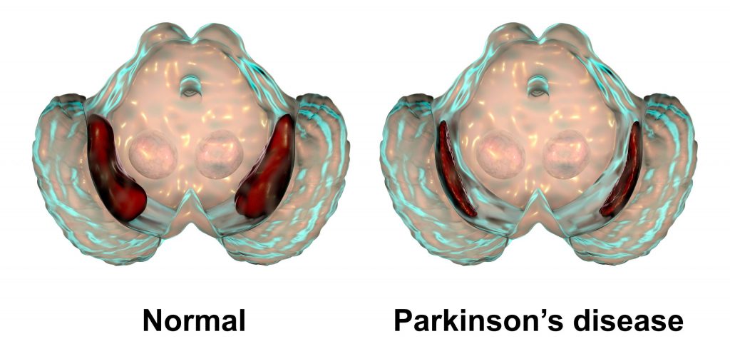
By definition, Parkinson’s Disease is a neurodegenerative disorder that affects predominantly dopamine-producing neurons in the Substantia Nigra (SN) (Latin for “Black Substance”, due to its darkened pigment in the brain). The substantia nigra contains the highest concentration of dopamine neurons. It is a part of the Basal Ganglia, an area responsible for motor control, motor learning, and procedural memory, such as learning how to tie your shoes. 

Substantia Nigra in Normal Brain vs. Parkinson’

Without the Substantia Nigra, the brain and body simply do not communicate as well, or at all depending on the stage of PD. To us, fitness professionals, we observe those living with Parkinson’s Disease displaying uncoordinated movements, loss of balance, poor gait, postural issues, visual tracking problems, rigidity, tremor, freezing, facial masking, inability to focus on a task or process information clearly, bradykinesia, hypophonia (volume of voice)… and as it pertains to Bilateral Coordination, the challenge to perform a drill with one side of the body such as the left arm while moving the right leg.  Now your wheels are turning and you are probably asking yourself…

Why Is Bilateral Coordination Important?  

Bilateral Coordination requires your small and large motor and visual-motor functioning to work together making it possible to accomplish ALL of your daily tasks! For example, in order to write your grocery list on a piece of paper, you need to hold the paper with one hand while writing the note at the same time. 

Proprioception or body awareness is another great reason to work on Bilateral Coordination. Throughout our entire life, we have the ability to know where our body is in time and space. For the person living with Parkinson’s Disease, this means a lower risk of falling, and believe me, that is crucial!

What Is The Scientific Explanation Of Bilateral Coordination?   

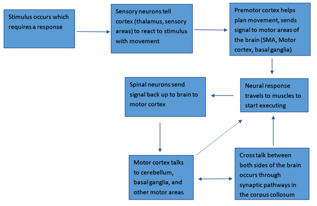
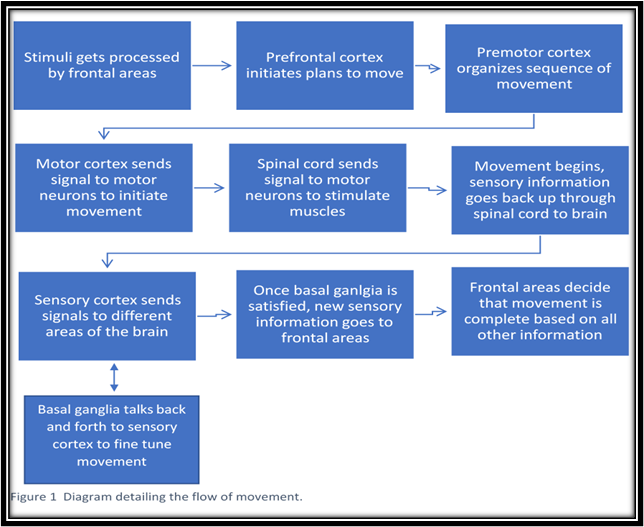
We learned in a previous article, “How a Thought Becomes an Action”, that motion in the brain is complicated and there are numerous steps to ensure that the motion in the brain is coordinated and precise. This all starts in the motor cortex, goes through the spine into motor neurons and muscles, and goes back into the brain to be fine-tuned by the basal ganglia (keep in mind this is where the substantia nigra resides). But now we’re adding onto this process by adding on even more coordinated movement in:

  • The premotor cortex
  • Supplementary motor area
  • The cerebellum
  • And the corpus callosum

Each one of these areas works in tandem with the movement pathways we discussed previously to produce bilateral movement and is the reason that I can type and you can read this paper right now. Let’s go over what each of these areas does, starting with the premotor cortex (Figure 1, below).

Fig. 1 The neural pathway from stimulus sensation to movement response.

  • The premotor cortex is more involved in the planning of movement rather than the execution is important for bilateral coordination in movements even as simple as walking. 
    • When walking, the premotor cortex would be important for planning the gait length required to keep our balance, the speed, and overall how the movement is going to go so we don’t crumple or trip over our own feet (to some extent anyway…)
  • The supplementary motor area (SMA) is also involved in planning movement, but in a different way. 
    • The SMA is planning for movements both ipsilaterally (scientific jargon for same-side), and contralaterally (scientific jargon for different-side).
      • While the premotor cortex is planning for what muscles have to move to do the actual action, the SMA is controlling which sides are going to go first to maintain balance and what makes the most sense given the environment we would be walking in.
  • The cerebellum is less involved in conscious movement but is extremely involved in coordination and balance, which is needed for performing tasks bilaterally. 
    • When walking, we need our brain to fine-tune the balancing and counterbalancing errors (such as arm swing), which happens in the cerebellum. Think of the cerebellum like an editor. The movement keeps happening, and each time it keeps getting better and easier because of the error calculations the cerebellum makes.
  • The corpus callosum, which works in tandem with the rest of the cortex to send signals to the different halves of the brain. 
    • What happens on one side doesn’t always translate to the other side, which is where the corpus callosum comes in. Each side has its own control mechanisms for the opposite side of the body (talk about contralateral). The corpus callosum is the phone line that connects these two hemispheres so they can talk to each other and get the messages for both sides of the body into a beautifully coordinated orchestra of neuronal firing and muscle movements.

So given these four areas of the brain, along with the rest of the movement pathway, there are numerous elements here contributing to sensory integration, planning of movement, checkpoints for these movement executions, and a miscommunication playing out on both sides of the brain. This means that there are a lot of alternate pathways in case anything goes wrong. However, pathways can deteriorate over time, and problems can arise. Which brings us to PD, and the issues that may happen in bilateral coordination as the disease progresses.

How Does Bilateral Coordination falter in the PD brain?

There’s a lot of different attributes to movements like walking –  a complicated movement with a number of variables:

  • First, you have to start walking.
    • Because it requires a lot of input all at once, it’s hard for the brain to do subconsciously, especially for someone who has PD. similar to revving a car for the first time after stopping at a red light.
  • Then you have sensory cues.
    • These can be challenges like incline or decline, which determines how much effort you need to stay upright, stay up to speed, and how much force you need to put into your step to keep up with gravity (like a driver constantly paying attention to the road).
  • Once you start walking, your brain can put less effort into what researchers call “steady-state walking”, where your legs follow the same instructions repeatedly, similar to coasting on a highway (2).
  • Next, we get to turning, where walking requires more input such as:
    • Balancing on one leg while pivoting the other
    • Slowing down, and speeding back up again (all while navigating those sensory cues, mind you), which is where a lot of the times this bilateral coordination seems to freeze up in PD, hence the clinical symptom called freezing of gait (FOG).
    • Thinking again of the car analogy, this is akin to having to slow down and turn the wheel the proper amount so you don’t hit the curb but you make it to the proper lane, speeding back up again.

With all those steps, anything that goes wrong will hurt the body’s ability to keep steady and coordinate larger and smaller movements on both sides. We also need to take into consideration that we all have a dominant side in normal movement that is easier to control. Evidence suggests the dominant side is worse in PD, perhaps due to brain asymmetry, meaning anything involving that side is most likely going to be slower and more uncoordinated, such as balancing on your dominant side or having to pivot on your dominant foot (1,2). Again, comparing it to a car running, if you have a bad steering wheel, your turns are going to be a little rough and you might end up swerving more or less on target. Or you need new adjustments or wheel bearings that make your balance and steadiness just a little bit better (speaking from personal experience). These parts make a car drive smoothly, as the neural circuits involved in the basal ganglia — along with all the new motor regions I mentioned–interact as one unit to make the walking as coordinated as possible.

This type of movement is also affected by our favorite neurotransmitter: dopamine. As we know, dopamine affects the basal ganglia, which has a conversation with all of the different areas we’ve talked about, such as the supplementary motor area, which is important for coordinating movements. We have seen in past research that taking dopaminergic medication has positively affected Phase Coordination Index (PCI), which measures bilateral coordination by looking at footsteps and foot switching (4). What does this mean exactly? Taking dopaminergic medications creates a more balanced movement, most likely by increasing basal ganglia activity, which thus increases the conversation between this and other movement areas needed for bilateral movement.

We also know that exercise is great for PD! Not only has this been seen in PD-specific fitness classes, but in research as well! Taking medications that increase dopamine uptake can mitigate some of these falters in movement and can create a more efficient signaling cascade, but it can only do so much. The other part of strengthening these pathways comes from exercise that can lead to better balance, smoother motion, and greater bilateral coordination! We’ve seen in some research studies on physical therapy techniques that auditory and visual cues, premeditated/thoughtful movements, and most importantly repeated balance drills can decrease FOG episodes, which could be attributed to a lack of bilateral coordination (3).

And what incorporates all of those? Exercise classes! In Part 2, we discuss Steps To Incorporating Bilateral Coordination Into Your Exercise Program.

Become a Parkinson’s Disease Fitness Specialist!

Check out Colleen’s online course on MedFit Classroom….


Co-Written by Colleen Bridges, M.Ed, NSCA-CPT and Renee Rouleau.

Colleen Bridges is the author of MedFit Classroom’s Parkinson’s Disease Fitness Specialist course. Renee Rouleau is a PhD student at the Jacobs School of Biomedical Sciences, University at Buffalo.


References

  1. van der Hoorn, A., Bartels, A. L., Leenders, K. L., & de Jong, B. M. (2011). Handedness and dominant side of symptoms in Parkinson’s disease. Parkinsonism & Related Disorders, 17(1), 58-60. https://doi.org/https://doi.org/10.1016/j.parkreldis.2010.10.002
  2. Plotnik, M., & Hausdorff, J. M.. (2008). The role of gait rhythmicity and bilateral coordination of stepping in the pathophysiology of freezing of gait in Parkinson’s disease. Movement Disorders, 23(S2), S444–S450. https://doi.org/10.1002/mds.21984
  3. Rutz, D. G., & Benninger, D. H.. (2020). Physical Therapy for Freezing of Gait and Gait Impairments in Parkinson Disease: A Systematic Review. PM&R, 12(11), 1140–1156. https://doi.org/10.1002/pmrj.12337
  4. Son, M., Han, S. H., Lyoo, C. H., Lim, J. A., Jeon, J., Hong, K.-B., & Park, H.. (2021). The effect of levodopa on bilateral coordination and gait asymmetry in Parkinson’s disease using inertial sensor. Npj Parkinson’s Disease, 7(1). https://doi.org/10.1038/s41531-021-00186-7
  5. Kramer P., & Hinojosa, J., (2010). Frames of Reference for Pediatric Occupational Therapy: 3rd Edition. Baltimore, Maryland: Lippincott Williams & Wilkins
  6. Magalhães, L.C., Koomar, J.A., Cermal, S.A. (1989, July) Bilateral Motor Coordination in 5- to 9-year old children: a pilot study. The American Journal of Occupational Therapy. Volume 43 Number 7.
  7. Piek, J.P., Dyck, M.J., Nieman, A., Anderson, M., Hay, D., Smith, L.M., McCoy, M., Hallmayer, J., (2003) The relationship between motor coordination, executive functioning and attention in school-aged children. Archives of clinical neuropsychology. Elsevier’s Ltd. doi:10.1016/j.acn.2003.12.007
  8. Roeber, B.J., Gunnar, M.R. and Pollak, S.D. (2014), Early deprivation impairs the development of balance and bilateral coordination. Dev Psychobiol, 56: 1110-1118. https://doi.org/10.1002/dev.21159
  9. Rutkowska, I., Lieberman, L. J., Bednarczuk, G., Molik, B., Kaźmierska-Kowalewska, K., Marszałek, J., & Gómez-Ruano, M.-Á. (2016). Bilateral Coordination of Children who are Blind. Perceptual and Motor Skills, 122(2), 595–609. https://doi.org/10.1177/0031512516636527
  10. Schmidt, M., Egger, F., & Conzelmann, A. (2015). Delayed Positive Effects of an Acute Bout of Coordinative Exercise on Children’s Attention. Perceptual and Motor Skills, 121(2), 431–446. https://doi.org/10.2466/22.06.PMS.121c22x1
  11. Tseng, Y., & Scholz, J. P. (2005). Unilateral vs. bilateral coordination of circle-drawing tasks. Acta Psychologica, 120(2), 172-198.
Senior-and-Trainer

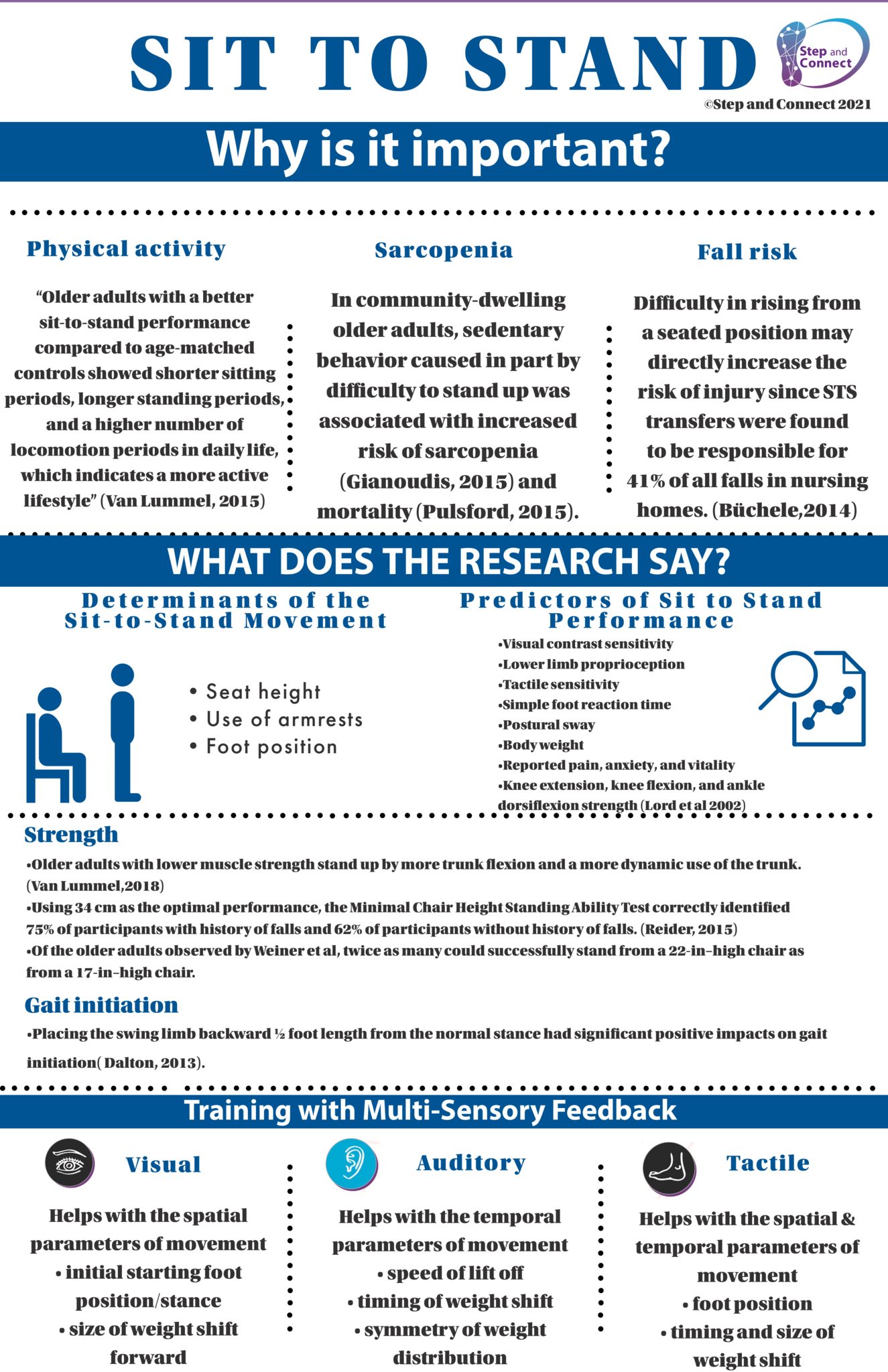
How Feedback Can Improve Sit to Stand Performance

Sit -to -stand transfers are important movements that physical therapists, occupational therapists, trainers, and coaches use every day with their clients. With professional guidance clients can successfully learn the symmetry of weight distribution and other mechanics required to correctly perform sit-to-stand. Without such guidance clients may not learn the safe and correct way to get up from a seated position or to use the movement as exercise. The result can be pain, falls and other injuries.

Feedback

Feedback regarding proper motor patterns is an important tool that can lead to greater movement efficiency, increased activity, and lower risk of injury. After analyzing the different phases of sit to stand (preparatory/ starting alignment, transitional movements, and the final standing posture), clinicians can use feedback to address both the spatial and temporal parameters of movement that is needed to improve overall performance.

Why We Need Feedback

A natural part of performing a skill is to use intrinsic feedback, the sensory perceptual information caused by-the movement. Vision, proprioception, touch, pressure, and audition help formulate a person’s internal representation of a movement goal.

When performing sit- to- stand, proprioceptors indicate the muscle length and tension of the position of the ankles and feet, as well as the amount of pressure through the limbs; visual information orients the individual to the environment; and vestibular inputs, contribute to sense of verticality. If your clients are not receiving the proper intrinsic feedback, they may not be aware of their movements. For example, an individual with impaired ankle proprioception may need extrinsic or augmented feedback to increase the weight and symmetry through their legs.

Augmented feedback enriches/enhances intrinsic feedback. It provides information to clients who are unaware of their body position. Augmented feedback can help engage the patient during all phases of sit -to- stand and with different modalities: visual, auditory or tactile.

What type of Feedback to Use? Auditory, Tactile, or Visual?

When training sit<>stand, what type of feedback and verbal cues would you provide to achieve forward weight shift, symmetry of weight distribution versus increasing speed of transfer?

Think about your clients who have trouble getting up from sitting and are not sure why they cannot rise on their first attempt. Often patients think the problem is lack of strength.

Sometimes using the cue “nose over toes” works for these patients. Other times, it is the size and timing of their forward weight shift that needs to be cued?

Auditory feedback provides an engaging solution in this situation. For example, the Step and Connect’s Balance Matters System features auditory feedback about the timing and amount of weight the client shifts forward. The system’s innovative foot pads make a clicking sound when the move is done correctly. The click is nonjudgmental and motivating. The client learns a new way to move without verbal instruction.  With practice the correct move becomes automatic.

During sit to stand transfers auditory feedback can improve: 

  • Starting alignment
  • Forward weight shift
  • Timing of weight shift
  • Symmetry of weight distribution
  • Postural control (decrease sway at the ankles)

Example using the Balance Matters system:

Sit to stand: Activate back clicker, activate front clicker while you stand, keep all clickers quiet during standing.

Stand to sit: Keep all clickers quiet until bottom is on the seat.

Progressions and Intervention Ideas

Part Practice (only one phase of the transfer).

  • Preparatory phase: Work on good starting posture, activating back clicker to promote anterior pelvic tilt and increased weight bearing through the legs.
  • Transitional phase: Reach forward with arms and activate front clicker to promote anterior weight shift.
  • Transitional phase for stand to sit: partial squats to sit down keeping clickers quiet to improve the timing of weight shift and decrease a “plop”.

Symmetry of movement and weight-bearing

  • Does one side activate sooner than another or do you not activate one clicker on one side since you are weight bearing more on the opposite side.
  • Verbal cue: Hear the clicker go off at the same time standing up and sitting down.

Activate the vestibular system standing on foam footpads

  • Add head turns with sit to stand on foam

Changing the Stance Position

  • Staggered stance and step to work on step initiation.

Eyes closed to improve balance in dimly lit environments.

  • This is important when standing up from bed to walk in order to go to the bathroom at night. An article “Effect of Sitting Pause Times on Balance After Supine to Standing Transfer in Dim Light” mentions that the risk of falling for older adults increases in dimly lit environments.
  • The results of the study suggest that longer sitting pause times may improve adaptability to dimly lit environments, contributing to improved postural stability and reduced risk of fall in older adult women when getting out of bed at night.
  • This is an important topic on how the speed or timing to adapt in different environments (dim lit or uneven surfaces) can change our overall balance and postural control and should be integrated into balance exercises and goals.

Using multi-sensory feedback with the Balance Matters system will:

  • Improve an individual’s awareness of their starting posture and transitional postures.
  • The auditory feedback in the footpads helps promote improved timing, sequence of the task, weight distribution for symmetry and weight shift.

Remember, there are influential factors when designing programs using feedback. Fading the feedback for retention is highly recommended. In the Balance Matters courses, we review more in-depth these influential factors and clinical applications.


Originally printed on stepandconnect.com. Reprinted with permission.

Balance Matters is a unique balance training sensory tool developed by recognized Physical Therapist Erica Demarch, intended initially for Parkinson’s patients and others suffering from balance issues. Now has become a popular sensory balance training tool for people looking to “train their brain” for improved balance. The Balance Matters system is the foundation of Erica’s company, Step and Connect, stepandconnect.com

senior-woman-lunge

How A Thought Becomes An Action: A Guide To Movement And The Disconnect In Parkinson’s Disease | PART 2

In Part 1, we discussed how a thought becomes an action, and the disconnect in Parkinson’s Disease, as well as how a Fitness Professional do to improve brain and body connection.

For those living with Parkinson’s, the three Activities of Daily Living (ADLs) considered to be most difficult to perform are:

  • Rolling over in bed
  • Getting out of a vehicle
  • Working through a freeze episode while crossing over a threshold between rooms.

I have provided a list of exercises to complement these ADLs as well as a “Practice Option” that combines the exercises listed.

Considerations

  1. Remember to begin with the most basic of movements until the client can properly and safely execute the exercise.
  2. Care partners of wheelchair-bound clients need to be instructed on how to safely assist loved ones without causing injury to either person. Please refer the client to an Occupational Therapist if needed.

Activity of Daily Living: Rolling over in bed                                         

Exercises

  • Bridges
  • Push-ups or chest press
  • Tricep extension
  • Rows
  • Glute squeezes (for chair bound)
  • Lateral Step with torso rotation using a tube
  • Side Planks/ Prone Plank
  • Clamshells

Advanced Practice Option: Have the client lay on his or her back. Take the right leg and swing the leg over the left leg and move into the side plank position and hold for 5 counts. From there roll to a prone plane OR bird dog position. Reverse the exercise to practice returning to the supine position.

Assisted Practice Option: If lying down is not an option, have the client sit in a chair.  Have the client hold a tube with both hands in front of them. Trainer provides tension from the side and the client maintains the isometric hold while picking up one leg and moving it out to the side and bringing it back in like a seated jumping jack.

Activity of Daily Living: Getting in/out of vehicle

Exercises

  • Rows (add a diagonal step/lunge)
  • Squats/Lunges
  • Sit to Stand drill (include single leg version)
  • Bridges
  • Clamshells (or any abduction work)
  • ½ Warrior step / ½ Gong arms
  • “Step over the Fence” ( lift left knee and step laterally over the “fence” followed by the right knee and then reverse the movement)
  • “Jazz Hands” (improves ability to reach)
  • Hip circles
  • Bob-n-weave (or lean left/right if needed)
  • Side planks or oblique bend
  • Tricep/Biceps (add lower body exercise)

Advanced Practice Option: (Stand with chair next to left leg). Place a hurdle next to chair to act as the “floorboard” of the car. Client will stand alongside the “car”.  Client will then lift the left knee and hold for 3-5 counts then step “over the fence”/bob-n-weave” to get into the car. Reverse the motion to practice getting out of the car. Repeat on the other side.

Assisted Practice Option: (Begin in a chair and with a short hurdle or object for them to “step” over). Client is in the chair and reaches right arm out as if opening the car door (jazz hands). Client then comes back to center and picks up the right knee and steps over the hurdle and turns foot to the right (½ warrior/ ½ gong) as the entire body turns to the right. Left foot follows the right foot and steps over the hurdle. Once the feet are facing the right, have the client do a full or partial Sit-to Stand drill. Reverse the motion to practice getting into the car.

Activity of Daily Living: Working through a “freeze” episode 

Exercises

  • Obstacle courses
  • Stop and start gait drills
    • Walk and turn head right and left
    • Walk slow then fast then slow etc
    • Walk and at cue, stop and turn
  • Visual drills
  • Lateral steps 5x then walk forward
  • Walk to a song with a strong beat
  • Criss-Cross Applesauce

*If client freezes at room threshold, emphasize that they want to look straight ahead and not down.

These three ADLs are just a few of the frustrating tasks people living with Parkinson’s Disease deal with each and every day.  Fitness Professionals can make a real difference in someone’s life if they will take the time to consider how movement works, where it can go wrong, and what to do to help it go right again. Imagine the success your client will experience during a session and throughout the day as they tackle ADLs with minimal effort! I can tell you this, their level of confidence will soar and the future will be something they look forward to.


Work with Parkinson’s Clients and Change Lives!

Working with Parkinson’s clients is an extremely rewarding experience. Check out Colleen’s course, Parkinson’s Disease Fitness Specialist to get started.


Colleen Bridges has worked for nearly 17 years as an NSCA Certified personal trainer, group exercise instructor and fitness consultant and as an independent contractor for Nashville’s first personal training center, STEPS Fitness. Her passion for understanding the body in sickness and in health, and how it moves, as fed her interest in and enhanced her talent for working with senior adults, especially those living with a neurological disorder such as Parkinson’s Disease.

Renee Rouleau is a Clinical Research Coordinator for the Department of Neurology at Vanderbilt Movement Disorder. Her research primarily focuses on the glymphatic system, a proposed waste-clearance system in the central nervous system in different neurodegenerative disorders such as Parkinson’s Disease (PD) and Alzheimer’s Disease (AD).

parkinsons-graphic

How A Thought Becomes An Action: A Guide To Movement And The Disconnect In Parkinson’s Disease | PART 1

It’s 2am and Robert needs to use the restroom but can’t gather enough strength to roll to a seated position to get out of bed without his wife’s help.

Gus decides to go to the kitchen for a snack but “freezes” when he reaches the doorway. His feet feel like they are stuck in mud.

Mary would like to attend her exercise class but the process of getting in and out of the car leaves her exhausted.

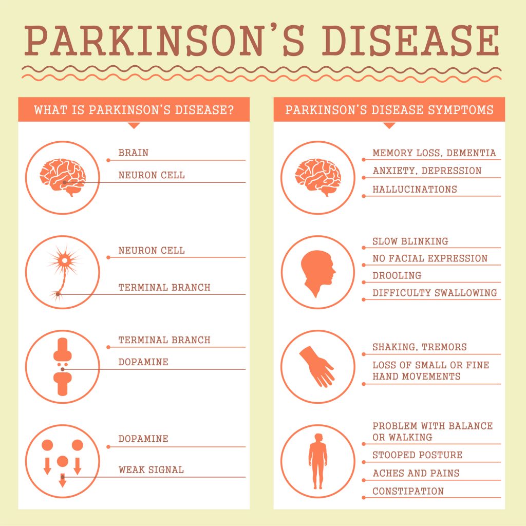
What do all of these people have in common? They have a progressive neurological disease called Parkinson’s Disease (PD). Parkinson’s Disease affects the dopamine-producing neurons in the substantia nigra (Latin for “Black Substance”, due to its darkened pigment in the brain).  The substantia nigra contains the highest concentration of dopamine neurons.  It is a part of the Basal Ganglia, an area that is responsible for motor control, motor learning, and procedural memory such as learning how to tie your shoes.

In PD, the onset of dopaminergic neuronal death in the substantia nigra manifests itself in the form of motor and non-motor symptoms that occur over a long period of time and in a progressive fashion.  Most people are not aware they are presenting symptoms of PD until a loved one brings their attention to a tremor, lack of arm swing, or notices a series of falls.

People living with Parkinson’s Disease want to take larger steps, smile more, swallow food without fear of choking, dress and bathe themselves, drive and participate in social activities.

However, for some, when they have a thought such as “I want to walk to the kitchen for a snack”, getting the thought to become an action, is almost impossible due to the lack of dopamine neurons in the Substantia Nigra. But wait a minute! HOW does a thought even become an action and WHAT can a Fitness Professional do to improve brain and body connection?

How a thought becomes an action

The brain is constantly multitasking as it takes in stimuli from your surroundings, interprets what’s going on around you and causes you to take action.  When your mind creates a conscious thought, such as “I want to get a snack”, a chain reaction takes place in the brain involving several areas. This starts in the frontal areas of your brain after processing the stimuli leading to the thought. For example, if you have your eyes set on the kitchen to get a snack, your prefrontal cortex initiates plans to make the movement, sending signals to your premotor cortex to organize those plans, and then sends those signals to the motor cortex to carry out the movement.

Once the movement has been planned and the best course of action has been “decided” by these neurons, the movement can commence. This creates the surge of neuronal firing from the motor cortex through the spinal cord to motor neurons that communicate with muscles and finally manifests the movement.

The above seems straightforward. The tricky part is regulating all of those different areas. Once the gross movement is executed, sensory information ( i.e. touch, temperature, or force) travels back up to the brain through sensory neurons in the spinal cord. The sensory cortex receives and carries the message to other parts of the brain that fine-tune the movement. This is one of the functions of the basal ganglia and other areas in the midbrain.

Because you’ve most likely done these types of movements before, those patterns are all stored in the basal ganglia so it doesn’t take up valuable space in the motor cortex. This area talks back and forth to the frontal areas to figure out what specific pattern should be used to achieve the best result. There are a hundred different ways to get out of a chair and go to the kitchen, but the basal ganglia works together to choose the most efficient option out of all of them and keeps the movements from getting out of control so you’re not high knee-ing to the kitchen when a simple walk will do (unless you want to high-knee to the kitchen). Once everything is adjusted and looks correct, new sensory information goes to the sensory cortex and back to those frontal areas to then signal that the movement has been fully executed.

Now, although that looked like a lot of steps just to complete one movement, this all happens within a fraction of a second, and is constantly going as you move to correct and adjust. The process is fluid, but works as a chain. If one link is broken, the rest of the process is going to fall apart. So how is the link broken in a disease like Parkinson’s?

Because the basal ganglia gets a lot of communication from the substantia nigra, if there is a loss of any sort of dopamine neurons, the relay of information gets discombobulated and, in the case of Parkinson’s, causes the motor system to stop the movement mid-way as there is not enough information from the neurons firing. Instead of creating the controlled movements and fine motor adjustments like you would see in a regular motor response, you have freeze-ups where the frontal areas are telling the midbrain to do one thing, and the basal ganglia just can’t do what it’s being told to do. Thus, the chain of movement is broken and the body cannot execute the action properly. To most, it looks like people with PD can’t seem to execute an action because of cognitive reasons. However, from their perspective, they want to be able to execute it and are consciously telling themselves to do it, but part of their brain isn’t “listening” and it causes the brain and body to be disconnected, resulting in incomplete movements and motor symptoms such as resting tremor, freezing of gait, and rigidity. This is why when PD patients take their medications, which help the brain to produce dopamine, they have “on” periods where these areas are able to have clearer communication with each other, their movements are better and their symptoms are better managed.

This is critical information for Fitness Professionals working with people living with Parkinson’s Disease. Once the information is understood, Fitness Professionals can focus on the second question which is “What can a Fitness Professional do to improve the brain-body connection for those living with Parkinson’s Disease?”

First, remind them that Exercise is Medicine! They need to take a dose each and every day! And the good news is that exercise provides outcomes such as:

  • Improved neuro-protection for at-risk dopamine neurons
  • Neuro-repair for areas of the brain affected by Parkinson’s Disease, and
  • Adaptation by retraining areas of the brain to pick up where the damaged parts can no longer execute commands.

Second, determine the activities of daily living (ADLs) that are most difficult for them. Identifying the ADLs and providing an exercise program that includes the seven functional movement patterns (push, pull, carry, hinge, lunge, squat and rotation) to improve their ability may save their lives. Repetition will be the key to create a spirit of confidence!

In Part 2, I discuss the three ADLs considered by most people living with Parkinson’s Disease to be most difficult, and exercises to complement them.


Colleen Bridges has worked for nearly 17 years as an NSCA Certified personal trainer, group exercise instructor and fitness consultant and as an independent contractor for Nashville’s first personal training center, STEPS Fitness. Her passion for understanding the body in sickness and in health, and how it moves, as fed her interest in and enhanced her talent for working with senior adults, especially those living with a neurological disorder such as Parkinson’s Disease.

Renee Rouleau is a Clinical Research Coordinator for the Department of Neurology at Vanderbilt Movement Disorder. Her research primarily focuses on the glymphatic system, a proposed waste-clearance system in the central nervous system in different neurodegenerative disorders such as Parkinson’s Disease (PD) and Alzheimer’s Disease (AD).

Senior-doctor-throat-open-wide

Parkinson’s Symptoms: “OPEN WIDE” – A Trip Down Our Throat

Our throat muscles, through which we speak, sing and scream, give us our signature sound. In many situations, people affected by Parkinsons disease (PD) have diminished voice control. According to Wikipedia, Parkinsons disease can cause changes in speech. The voice may get softer, breathy or hoarse, causing others difficulty hearing what we say. Speech may be slurred. Speech changes can interfere with communication, which can be isolating and harmful.

Other causes of voice disorders include infections, stomach acids that move upward in the throat, growths due to a virus, cancer and diseases that paralyze the vocal cords. Here is a brief understanding of what, how and ways to care for your precious voice.

What Are Your Vocal Cords?

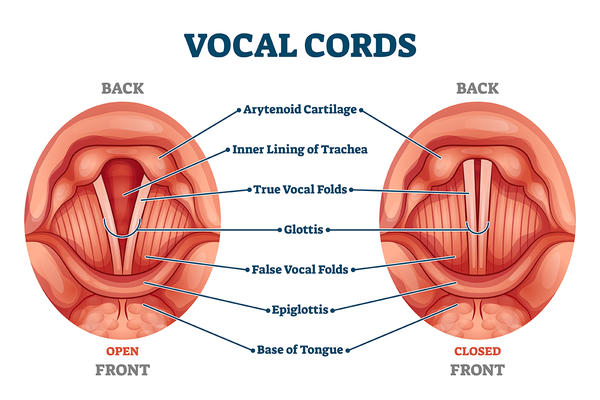
Your voice box sits between the base of your tongue and the top of your windpipe, which is where your Adams apple likes to hang out. (The Adams apple is more pronounced in men than in women.) The vocal cords are two bands of smooth muscle membrane tissue, each covered in a mucous membrane, that stretch across the voice box like the strings on a guitar.

How They Work

When youre quiet: listening, observing, perhaps meditating your vocal cords sit apart, creating a tunnel through which you breathe in. But the moment you begin speaking, they clap together as the diaphragm pushes air up from the lungs. This air causes a buzz sound or vibration and sends sound waves through your throat, nose and mouth, which amplify them. As these humspass through, they are transformed into song or sentences.

Your Sound

Vocal cords vary in thickness and length, which is why each person has his or her unique tune. Think of the singer Barry Whites voice. Deep and strong. Those who have booming voices, such as Barbra Streisand have larger resonating cavities (throat, nose and mouth).

Take Care of Your Cords

Yelling, or screaming can cause inflammation and lesions on your vocal cords. Even a long-lasting cough can do damage. If you are hoarse, rest your voice. Speak softly and try to avoid throat clearing, even if your throat is congested. Drink lots of water to thin excess mucus and lubricate. Menthol and eucalyptus can be irritating.

For those with Parkinsons disease, if you notice your voice is diminished, see a speech-language pathologist (SLPs) specializing in voice therapy. They can assist with diagnosis, assessment, planning and treatment of voice disorders including difficulty with swallowing. Some exercises include moving your tongue up and down, moving it from corner to corner, placing your tongue at the tip of your mouth or smiling and saying EEEloudly. Repeat 10 times, at least twice a day.


Reprinted with permission from Lori Michiel. 

Lori Michiel, NASM, has been assisting seniors in their homes since 2006 with customized exercise programs including those designed to address Parkinson’s, metabolic disorders, arthritis and diabetes. These adaptive programs are specifically designed to improve balance, circulation, flexibility, mobility and promote independence. Lori Michiel Fitness has over 40 certified trainers who are matched with clients in Los Angeles, Ventura and Orange Counties. Connect with Lori at www.LoriMichielFitness.com.

brain-neurons

Parkinson’s Disease and Exercise

Parkinson’s disease is the second most common neurodegenerative disorder after Alzheimer’s disease.  Unfortunately, the incidence of Parkinson’s disease has not declined, and its impact is seen in all races.  This is due in part to the fact that the population of the world is greater than ever before and increasing. In addition, people are living longer than in previous generations, and the baby boomer generation, one of the largest generations in history, has reached old age.

Risk factors for Parkinson’s disease include:

Age: Risk of Parkinson’s disease increases with age.  The average age of onset for this disease is 55 years and the rate of incidence increases steadily until the age of 90.

Gender: Men have a higher risk for Parkinson’s disease than women.

Family history: Individuals with a family history of Parkinson’s disease are at a higher risk for Parkinson’s disease. Moreover, it is said that those with affected first-degree relatives double their risk of Parkinson’s disease.

Agricultural work: Individuals exposed to pesticides and herbicides have a greater risk of developing Parkinson’s disease symptoms. Drinking well-water and living in rural areas have also been associated with an increased incidence of Parkinson’s disease.

Head Trauma: Head trauma can be a risk factor for Parkinson’s disease as is seen in the case of boxers. One study showed that trauma to the upper cervical region, head, and neck was a risk factor for Parkinson’s disease. However, in some cases it took years for these symptoms to appear.

The exact cause of Parkinson’s disease is unknown.  Regarding the molecular events that lead to the development of this disease, there is still some uncertainty in terms of what causes the neurodegeneration seen in Parkinson’s disease. The current hypothesis is that Parkinson’s disease may result from the interaction between environmental factors and genetic susceptibility.

The primary symptoms for PD are deficiencies in motor performance due to the loss of the dopamine pathways in the brain. Decreased dopamine production in the substantia nigra in the brain causes the 4 primary motor symptoms:

  • Bradykinesia: described as slowness in the execution of movements while performing daily activities.
  • Rigidity or Stiffness: caused by an involuntary increase in tone of the limbs and axial musculature.
  • Resting Tremor: Found primarily in the arms and hands and can be socially bothersome. Resting tremors are less disabling since they often vanish with the initiation of activity (especially in the early stages of Parkinson’s disease).
  • Postural Instability: manifested in a slow speed of walking, shortened stride length, narrowing of base of support, and leaning towards one side.

Exercise should be targeted for the primary motor symptoms with exercise and occupational therapy to improve quality of life. Recommended program components include:

  • Posture, gait, mobility
  • Fall risk reduction
  • Cardiorespiratory health
  • Strength and function
  • Depression and Anxiety
  • Joint health

Exercise prescription for clients with PD includes: (ACSM)

  • An individualized program
  • Cardiorespiratory: use guidelines for healthy adults
  • Muscular Fitness: use guidelines for healthy adults
  • Flexibility: slow, static exercises for all major and minor joints in the body including the upper torso, spine, and neck.
  • Neuromotor Exercises: help with balance, gait, and postural instability. Clinicians use a gait belt or parallel bars to ensure safety depending on the severity of the symptoms.  Include functional exercises to improve ADLs and quality of life.

PD exercise therapy includes intervention with many kinds of exercise modes. Both personal training and group fitness have been successful in helping to manage the disease and reduce the symptoms. There is not strong evidence at this point to show that exercise prevents PD, but it is believed that exercise may play a role.  Exercise is however the mainstay for symptom management and slowing disease development.


June M. Chewning BS, MA has been in the fitness industry since 1978 serving as a physical education teacher, group fitness instructor, personal trainer, gym owner, master trainer, adjunct college professor, curriculum formatter and developer, and education consultant. She is the education specialist at Fitness Learning Systems, a continuing education company.

References and Resources:

brain

Neuroplasticity and the Aging Brain

One of the greatest concerns for the aging population is cognitive decline which leads to loss of independence as well as an extreme burden on the caretakers.  Individuals worldwide are fearful of being diagnosed with any of the various cognitive issues: Dementia, Parkinson’s, Alzheimer’s, and other forms of cognitive debilities.  In 2015 there was an estimated 47 million people living with dementia and this number is expected to triple by 2050.  In 2014, the Alzheimer’s Association reported that they believe there is sufficient evidence to support the link between several modifiable risk factors and a reduced risk for cognitive decline and sufficient evidence to suggest that some modifiable risk factors may be associated with reduced risk of dementia. Specifically, that regular physical activity and management of cardiovascular risk factors (diabetes, obesity, smoking, and hypertension) reduce the risk of cognitive decline and may reduce the risk of dementia. The Association also believes there is sufficiently strong evidence to conclude that a healthy diet and lifelong learning/cognitive training may also reduce the risk of cognitive decline.

Positive association between aerobic exercise or CV fitness and executive functions is highly consistent but cannot determine causality.  Aerobic exercise (AE) has shown moderate to medium sized effects on executive function and memory. Resistance Training (RT) has improved executive function and memory. Combined AE and RT has the biggest (potentially synergistic) effect. It has been proposed that the physical and cognitive exercise might interact to induce larger functional benefits.  Larger benefits on cognitive test performance were noted for combined physical and cognitive activity than for each activity alone.  “Claims promoting brain games are frequently exaggerated and at times misleading. … To date, there is little evidence that playing brain games improves underlying broad cognitive abilities, or that it enables one to better navigate a complex realm of everyday life (Consensus statement, 2014).

Neuroplasticity is the brain’s ability to reorganize and rebuild itself by forming new neural connections. The more neural pathways you have, the more resilient your brain is. Neurogenesis is the process of creating new neurons (brain cells).

Contrary to popular belief, neurogenesis continuously occurs in the adult brain under the right conditions such as with exercise.  Substantial benefits on cognitive test performance were noted for combined physical and cognitive activity than for each activity alone. It was also noted that the physical and cognitive exercise together might interact to induce larger functional benefits.  “We assume, that physical exercise increases the potential for neurogenesis and synaptogenesis while cognitive exercise guides it to induce positive plastic change” (Bamidis, 2014).  To maximize cognitive improvement, combine physical exercise with cognitive challenges in a rich sensorimotor environment that includes social interaction and a heaping dose of fun.

Brain health is becoming extremely important as individuals live longer.  Today there is much more information available on how to train the aging brain.

Some great resources are:


Dianne McCaughey Ph.D. is an award winning fitness specialist with more than 35 years experience in personal training, group exercise, coaching, and post-rehabilitation. She is a master trainer for multiple companies and practices and teaches optimal wellness emphasizing the mind, body and spirit. She works with special populations and focuses on posture, gait, balance and corrective exercise programs for better function and health.

Cody Sipe, PhD, has an extensive background in the fitness industry with 20 years of experience as a personal trainer, fitness instructor, program director, exercise physiologist and club owner. He is currently an Associate Professor and Director of Clinical Research in the physical therapy program at Harding University. He is the co-founder and vice president of the Functional Aging Institute (FAI).