Hide

Error message here!

Lost your password? Please enter your email address. You will receive a link to create a new password.

Error message here!

Back to log-in

Close
hand xray bones

7 Simple Habits You Didn’t Know Could Affect Your Bones

Do you know that the things you do every day can affect your bones? Sadly, ‘harmless’ things that are a part of our routine can actually hurt our bones in the long run. For example, we carry a lot of weight, we lead a sedentary life, we eat all the wrong things; and with the passage of time, we develop joint aches and bone ailments.

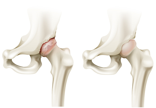
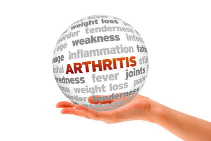
By eliminating these simple habits from our lifestyles, we can avoid getting osteoarthritis and protect our health.  Continue reading to find out more.

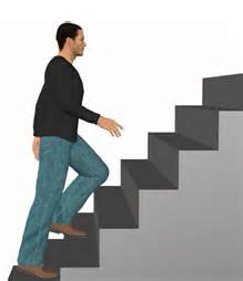
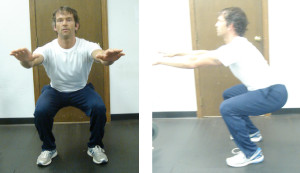
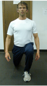
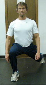
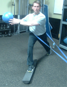
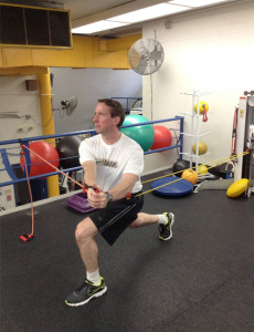
1.     Sedentary Lifestyle

We wake up, go to work, and sit in front of a computer screen all day. People who spend a lot of time sitting have at a higher risk of suffering from osteoporosis. Practicing a sedentary lifestyle is common for people who work in an office. There is not much time to move around.

Walking, dancing and running can be very helpful in strengthening your bones. So give your bones the exercise they need.

2.     Lack of Sleep

Rest is essential for everyone. The thing you should know about your body is that it goes through a constant cycle of detoxifying and cleansing. This process starts with the lymphatic system and continues to your liver. They slowly re-balance and restore the essential functions of your body. And for this process to occur, your body needs to be submerged in deep rest.

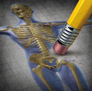
Research shows that lack of sleep damages the health of your bones as well. It also reduces the bone marrow and makes materialization difficult, which can lead to osteoporosis in the future.

3.     Be Careful with High Heels and Bags

Women have a lot of habits that damage their bones over time. We all drool over our high heels and spend a considerable portion of our paycheck buying the perfect pair. What we don’t know that wearing high heels frequently have a terrible impact on our posture. And thereby, affect the bones in our shins, feet, and back. After a long day of wearing heels, intense pain and fatigue sets in. And in the years that come by, you could suffer from serious problems. So instead of choosing high heels, go for the medium ones.

Another thing that is affecting your posture is those heavy bags. Without even realizing it, some women carry 10 kilos worth of stuff in their tote and shoulder bags. Carrying so much weight, unfortunately, puts a great deal of stress on your shoulders and spine. Not to forget, it causes pain and discomfort.

4.     Smoking

We have all heard the warning that smoking is bad for health. What most people don’t know is that it is even worse for your bones. Studies prove that people who use tobacco frequently have lower levels of bone density. Smokers are at a higher risk for fractures than non-smokers. Smoking produces free radicals which kill the cells that build bones (i.e., osteoblasts). If you have already had a fractured bone, smoking damages your blood vessels, which result in the slow healing process.

So throw away that pack of cigarettes, you don’t need them, and neither do your bones.

5.     High Salt Consumption

There is definitely a relationship between lower bone density and high salt intake.

We all know salt is bad for our skeletal system as it leeches calcium from our bones. The junk food that we like to munch on time and again is also causing harm to our joints and ligaments. Table salt consists of chloride and sodium. If we consume too much of it, it promotes metabolic acidosis. And that contributes towards the loss of bone density in the long run.

6.     Constant Coffee and Soda Consumption

Your day isn’t complete without a cup of coffee. Caffeine is necessary to wake up and give our day the much-needed boost. Coffee contains Methylated xanthine, which increases the amount of calcium release through urine. And over time, the minerals from your bones are affected, resulting in brittle bones.

One or two cups of coffee a day is fine. But, if you exceed that number, your bones will suffer the consequences.

Another drink that affects the health of your bones is soda, especially cola drinks. Sodas have a high amount of phosphoric acids in them, which reduce the consumption of calcium. So it’s better to avoid carbonated beverages so they can’t cause problems later.

7.     Sugary or Processed Foods

Packaged and highly-processed foods have low nutrients and high sodium and sugar level. Processed food contains food additives and synthetic chemicals that are very bad for your health. Packaged foods often include hydrogenated oils that are very damaging to your bones. In addition to that, your bones will pay a hefty price if you indulge yourself in a lot of sugary snacks. Not only sugar is inflammatory; it also leads to the blood-sugar imbalances that damage your bones.

Remember, if you cut back on these 7 habits, your bones will be stronger, and you will avoid a lot of bone-related problems in your later life. Not to forget, watch your calcium and vitamin D intake so your bones could be healthy!


Zyana Morris is a passionate blogger who loves to write on trending health, fitness and lifestyle topics. She is a featured author at HealthableUplifting FamiliesInscriber Mag, Hello Mamas and few others. Her favorite quote “It does not matter how slowly you go as long as you do not stop”. You can follow her through Facebook and Twitter.

ORTHOPEDICS CONSULTATION WOMAN

Rebuilding and Regenerating Damaged Knees: The Future Has Arrived!

If you grew up in the 1970s, you may remember the Six Million Dollar Man TV show with Steve Austin. The show began with a futuristic-type regeneration of an injured Steve Austin, and they would say, “we have the technology, we can rebuild him.” It was a great one-liner, but seem so far-fetched. Fast forward 30 years, or just one generation. The future is now. We have the technology!

gellert-hip1

The Hip Complex: Understanding the Science Behind Both Movement and Dysfunction

Introduction

The foot is where movement begins requiring mobility to initiate daily and sport specific movements. However, the knee however, requires stability with daily movements, but more importantly, dynamic sport movements such as soccer or football. The hip, like the ankle, requires mobility, to perform such simple movements as sit to stand, climbing stairs and other functional movements. In this article, we will review the anatomy of the hip, common injuries to the hip, functional assessments and training strategies to work with clients with previous injuries.

Figure 2. Hip joint with supporting ligaments

Hip joint with supporting ligaments

Let’s look at the basic anatomy of the hip. The hip joint is a multi-axial ball and socket joint between the femoral head and the acetabulum, similarly to the shoulder joint. The hip is surrounded in white, several ligaments that provide support and stability.

The labrum attaches to the acetabulum deep within the socket between the femur and acetabulum. The joint is covered by a capsule blended with three strong ligaments: iliofemoral or “Y” ligament, which resists extension, the ischiofemoral ligament, which resists extension and internal rotation, and the pubofemoral ligament, which resists abduction.

Muscularly, the glute medius and minimus are located along the anterolateral aspect of the hip to stabilize in the frontal pane, whereas the glute maximus, is located in the sagittal plane posteriorly to facilitate hip extension.

gellert-hip3

Supporting muscles around the hip

Common injuries and causes

There are different types of injuries the hip can sustain. The most common are the hip osteoarthritis, iliotibial band syndrome and total hip replacement. In this next section, we will review each condition providing a deeper understanding of each.

gellert-hip4

Osteoarthritic hip on left, normal hip on right

a. Hip osteoarthritis(OA)

Mechanism of injury/pathophysiology: A degenerative process of varied etiology which includes mechanical changes within the joint. O.A. affects some 40% of those aged over 65 in the community may have symptomatic OA of the knee or hip(Zhang, W. et al 2007).

Pathophysiology: Osteoarthritis (OA) is a relatively common musculoskeletal disorder, with a high prevalence that increases with age. O.A. is a degenerative process of varied etiology, which includes mechanical changes within the joint (Pisters, M., et al 2007).

Risk Factors: Excessive weight born on hip joint, muscle imbalance, repetitive stressors.

Sign and symptoms: Pain in the a.m. described as “achy” that decreases as the day progresses, pain with weight bearing or walking, difficulty squatting, and lateral thigh discomfort. Patients will describe of pain and stiffness in the a.m. described as “achy.” During the day, movement and activity, improves mobility and activity(Fernandes, L et al 2010). However, the volume of activity if too much, will increase pain. Patients typically have pain with weight bearing or prolonged walking, difficulty squatting, and lateral thigh discomfort.

Medical treatment: Non steroidal (NSAIDS)(examples are Ibuprofen/Advil).

gellert-hip5

Iliotibial band syndrome

b. Iliotibial band syndrome(ITB)

Mechanism of injury: Iliotibial band syndrome (ITBS) is a common injury of the lateral(outside) aspect of the knee particularly in runners, cyclists and endurance sports. ITBS is the most common running injury(Ellis, R et al 2007).

Pathophysiology: ITB syndrome is a non-traumatic overuse injury caused by repetitive friction/rubbing of the distal(farthest) portion of the iliotibial band (ITB) over the lateral femoral epicondyle with repeated flexion and extension of the knee.

Contributing/Risk Factors:
• Muscle imbalances/weakness: per the research and my clinical experience, hip flexors and quadriceps are stronger and than the hamstrings.
• Shoe support-important to rotate running shoes every 6 months or 500 miles according to multiple podiatrists I have worked with over the years.
• Increased bouts of running, altered foot mechanics-ie. Orthotics or need for orthotics.
• Lack of stretching, particularly tight ITB, hip flexors and quadriceps. Contributes to increasing compression along the outer hip.

Sign and symptoms: Lateral knee pain over the lateral condyle of the femur described as “dull/achy” that gradually develops & worsens particularly with running. Pain then becomes “sharp” in nature.

c. Total hip replacement

Mechanism of injury: Osteoarthritis is a musculoskeletal condition that develops over time affecting primarily the hip and knee joints. O.A. affects some 40% of those aged over 65 in the community may have symptomatic OA of the knee or hip(Zhang, W. et al 2007).

Pathophysiology: Osteoarthritis (OA) is a relatively common musculoskeletal disorder, with a high prevalence that increases with age. O.A. is a degenerative process of varied etiology, which includes mechanical changes within the joint (Pisters, M., et al 2007). Significant pain, decreased mobility and compromised function, are the primary reasons, a person would typically undergo a total joint arthoplasty(joint replacement). Total joint arthroplasty is a highly efficacious and cost-effective procedure for moderate to severe arthritis in the hip(Santaguida, P. et al 2008).

Common assessments

A simple functional test to assess a client’s movement pattern, is the squat. The squat is a classic fundamental primal movement that someone typically performs almost on a daily basis. With this test, you can observe how the client’s ankle, knee, hip and back moves compared to normal movement patterns.

What am I looking for?
The approach to assessment is all about asking and answering questions about movement:
• How does the client start, finish the movement?
• What strategies do they use? Do they have the appropriate flexibility to perform the movement?
• Is stability a problem? Are there compensations elsewhere in the movement sequence?

How do I interpret the movement?
• It is important to observe the client in both the frontal and sagittal planes
• Observing globally first, then examine how the entire kinematic chain is working as it relates to timing and sequence to achieve the movement

Dynamic Movement Assessments

1. Functional squat
The squat is a classic fundamental primal movement that someone typically performs almost on a daily basis. Whether it is to perform to pick something up or move an item. Therefore, it is important to assess the movement pattern a client uses during this movement.

gellert-hip6

Squat in frontal view; Squat in side view

What is required in a squat?
• Adequate ankle mobility, knee stability, hip mobility, lumbar spine(lumbo-pelvic junction) stability

gellert-hip7

In place lunge

Observations:
• Note the overall quality and range of movement in the frontal plane and sagittal plane
• Note the symmetry or lack of symmetry with the movement
• Note the point of transition from descending to ascending
• Note if there is an shaking(juttering), which indicates weakness in the lumbo-pelvic junction affecting the entire kinematic chain

Another simple assessment is an in place lunge, which examines one’s control through the entire kinematic chain. The lunge is another fundamental primal movement. The lunge is a dynamic movement that is typically performed during daily activities (stooping down to pick something up) or as part of an athletic movement. This test examines ankle control, knee control and pelvic movement in the sagittal plane.

Training strategies and programming for hip injuries

With any injury, the most important thing to remember is the type of injury, healing time and prior level of function of the client. Let’s begin with ankle sprains.

gellert-hip8

SLS stance with TRX

a. Hip Osteoarthritis(O.A.)

gellert-hip10

ITB stretch

Recommendations for training: Aqua therapy has been shown in the research to significantly reduce pain, improved physical function, strength, and quality of life(Hinman, Rana S., et al 2007), stretching ITB, hip flexors, quadriceps and hamstrings, strengthening weaker hip abductors(glute medius/minimus). Strengthening specifically hip abductors in various studies when compared to general strengthening, resulted in significant reduction in knee pain, objective change in functional outcome tests, physical function and daily activities(Bennell, K.L., et al. 2010 & Hernández-Molina, G et al. 2008). Core strengthening should also be an integral part of the training program.

b. ITB Syndrome

Recommendations for training: Important to keep stretching the ITB after exercise. Client education on the changing of running shoes every 500 miles or 6 months is key. Resistance training should focus on strengthening weaker phasic muscles (glute medius, minimus and maximus), which are required to stabilize and push off during running. Dynamic core strengthening should always play an integral role of training. Use of aqua therapy can be extremely beneficial and relaxing. It is very important to educate the client on the importance of cross training(ie. yoga, pilates, hiking, and swimming to condition the lower extremity muscles.

c. Hip replacement(THR)

Bridging with physioball

Bridging with physioball

Recommendations for training: Prior to commencing training, it is important to clarify with client, that there are no other underlying health issues. Important to follow hip precautions: avoidance of crossing affected leg towards midline(adduction), and squatting past hip flexion 90 degrees. Training should focus on strengthening weak glute medius/minimus, glute maximus and hamstrings. Effective and safe exercises include; in place lunges, diagonal lunges, seated leg extension and seated leg curl machine. Core strengthening should also always begin with static exercises, and then progressed to dynamic accordingly. Safe and effective core strengthening exercises include; standing trunk rotation with tubing or cable, four point planks, and side planks. Safe dynamic core strengthening exercises include bridging with physioball, single leg bridge with physioball, traveling forward lunge with medicine ball trunk rotation, and four-point plank on physioball as examples.

Summary

The hip is a complex unit thitbdat is comprised of a multitude of ligaments, tendons, connective tissue, muscles that synergistically initiate and correct movement, and stabilize when an unstable environment. Understanding the anatomy, biomechanics and weak links of the hip, common injuries and evidenced based training strategies, should provide you with the insight to better understand and work with clients with these kind of injuries more confidently.


Written by Chris Gellert, PT, MMusc & Sportsphysio, MPT, CSCS, AMS.

Chris is the CEO of Pinnacle Training & Consulting Systems (PTCS). A continuing education company, that provides educational material in the forms of home study courses, live seminars, DVDs, webinars, articles and min books teaching in-depth, the foundation science, functional assessments and practical application behind Human Movement, that is evidenced based. Chris is both a dynamic physical therapist with 14 years experience, and a personal trainer with 17 years experience, with advanced training, has created over 10 courses, is an experienced international fitness presenter, writes for various websites and international publications, consults and teaches seminars on human movement.

REFERENCES
Bennell, K.L., et al. 2010, ‘Hip strengthening reduces symptoms but not knee load in people with medial knee osteoarthritis and varus malalignment: a randomized controlled trial,’ Osteoarthritis and Cartilage, vol. 18, issue 5, pp. 621–628.

Ellis, R., et al., 2007, ‘Iliotibial band friction syndrome—A systematic review,’ Manual Therapy, vol. 12, pp. 200–208.

Fernandes, L., et al 2010, ‘Efficacy of patient education and supervised exercise vs. patient education alone in patients with hip osteoarthritis: a single blind randomized clinical trial,’ Osteoarthritis and Cartilage, vol. 18, issue 10, pp. 1237–1243.

Goodman, Catherine., Boissonnault, William., 1998, G. Pathology: Implications for the Physical Therapist, W.B. Saunders Company, Philadelphia, pp. 267-274, 279-292, 318-328, 412- 417, 609-610, 614-615, 617-621, 660-667, 736-745, 748-755.

Hernández-Molina, G et al. 2008 ‘Effect of therapeutic exercise for hip osteoarthritis pain: Results of a meta-analysis,’Arthritis Care & Research, vol. 59, issue 9, pp. 1221-1228.

Hinman, Rana S., et al 2007, ‘ Aquatic Physical Therapy for Hip and Knee Osteoarthritis: Results of a Single-Blind Randomized Controlled Trial,’ Journal of Physical Therapy, vol. 87, no. 1, pp. 32-43.

Pisters, M., et al., 2007, ‘Exercise adherence improving long-term patient outcome in patients with osteoarthritis of the hip and/or knee, Arthritis Care & Research, vol. 62,

Santaguida, P. et al 2008, ‘Patient characteristics affecting the prognosis of total hip and knee joint arthroplasty: a systematic review,’ Canadian Journal of Surgery, vol. 51, issue 6, pp. 428-436.

Zhang, W., et al., ‘OARSI recommendations for the management of hip and knee osteoarthritis, Part I: Critical appraisal of existing treatment guidelines and systematic review of current research evidence,’ Osteoarthritis and Cartilage, vol. 15, issue 9 pp. 981–1000.

Doctor Examining Male Patient With Knee Pain

The knee complex: understanding the science behind both movement and dysfunction

Introduction

The foot is where movement begins, requiring mobility to perform simple functional movements. The knee however, requires stability with daily movements, but more importantly, dynamic sport movements such as soccer or football. In this article, we will review the anatomy of the knee, common injuries of the knee, functional assessments and training strategies to work with clients with previous injuries.

Basic anatomy of the knee
Let’s look at the anatomy of the knee.. The joint is vulnerable when it comes to injury, because of the mechanical demands placed upon it and the reliance for soft tissue to support the knee. There are two primary joints within the knee, the tibiofemoral joint and the patellofemoral joint.

Figure 2. Structures within the knee joint

Figure 2. Structures within the knee joint

Knee Joints

a. Tibiofemoral joint: Is a hinge joint that permits some rotation between the distal end of the femur and proximal end of tibia. The joint capsule surrounds the femoral condyles and tibial plateaus and provides stability to the knee by the medial collateral ligament(MCL) and the lateral collateral ligament (LCL).

b. Patellofemoral joint: Is formed by the patella(knee bone) that glides in the trochlear groove of the femur. The height of the lateral femoral condyle helps prevent lateral subluxation, while soft tissue surrounds the joint to increase stability. This is seen in figure 3.

Figure 3. Patellofemoral joint

Figure 3. Patellofemoral joint

Primary structures within the knee joint: ligaments and mensici
Several ligaments described below provide stability at the knee joint.

a. Collateral ligaments: The two primary supporting ligaments are the medial collateral ligament (MCL), which is along the inside of the knee. The MCL is a thinner and weaker ligament biomechanically, making it more susceptible to injury more often injured per the research. While the lateral collateral ligament(LCL) is along the outside or lateral aspect of the knee providing lateral knee stability.

b. Anterior cruciate ligament(ACL): is the most commonly injured knee ligament and is taut during knee extension. It originates more proximally on the femoral side than the posterolateral (PL) bundle. It inserts anteromedially(front and to inner side) on the tibia. The ACL limits and controls forward translation of tibia on the femur and limits tibial rotation.

c. Menisci: the menisci are fibro cartilaginous discs located on the articular surface of the tibia along the medial and lateral tibial plateaus. The outer portion of the meniscus(lateral meniscus)is oval shaped (O) and thick. Attaching at the anterior and posterior horns via coronary ligaments.

Vascularity: The middle third and inner third of both menisci are relative avascular. The medial meniscus is more C-shaped, and thinner in structure. Both menisci receive nutrition through synovial diffusion and from blood supply to the horns of the menisci.

Function of the menisci: The menisci provide shock absorption, joint lubrication and stabilization.

Common injuries and causes

There are several common injuries that affect the knee. The most common are patella femoral syndrome(PFS), osteoarthritis(O.A.) and anterior cruciate ligament(ACL) injuries.

In this next section, we will review each condition providing a deeper understanding of each.

a. Patellofemoral syndrome
Pathophysiology/sign and symptoms: PFS is a condition where the patella does not translate biomechanically in the trochlear groove between the femoral condyles. Here the patella is positioned in either a tilt, glide or rotation accompanied by diffuse, achiness in the front of the knee.

Contributing Factors(Evidence Based Research): Several studies have shown that decreased flexibility of quadriceps and hip flexors(Lankhorst et al. 2012 & Meira et al. 2011) contribute to PFS. Decreased hip abductor strength has been shown a significant factor seen in multiple studies as contributing to PFS (Khayambashi, H., et al. 2012, Meira et al, (2011), Bolgla et al. (2008),Cichanowski et al. (2007), and Robinson et al. (2007).

Other factors include prolonged wearing of high heels, muscle imbalances(quadriceps>hamstrings).

b. Osteoarthritis(OA) of the knee

Figure 4. Osteoarthritis of knee

Figure 4. Osteoarthritis of knee

Pathophysiology/sign and symptoms: A degenerative process of varied etiology, which includes mechanical changes within the joint as seen in figure 4.

Risk Factors: Excessive weight born on hip joint, muscle imbalance, repetitive stressors.

Sign and symptoms: Pain in the a.m. described as “achy” that decreases as the day progresses, pain with weight bearing or walking, difficulty squatting, and lateral thigh discomfort.

c. Anterior cruciate ligament injuries
In the last several years, there has been more news about the incidence of ACL injuries. The incidence rate is greatest between the ages of 16 and 18 years. Female athletes are 3-9x more likely to sustain an ACL injury then male athletes. This results in at least 200,000 ACL reconstructions are performed each year in the United States, with estimated direct costs of $3 billion (in U.S. dollars) annually (Frobell, R., et al 2010).

Figure 5. Mechanism of injury for ACL tear

Figure 5. Mechanism of injury for ACL tear

Pathophysiology/Mechanism of Injury: The knee is struck while in hyperextension, forcing tibia anterior(forward)on the femur, as seen in figure 5. The ACL can also be injured with same mechanism of injury with combined with medial rotation of the lower extremity(LE). This creates instability and a direct disconnect the nervous system to the musculoskeletal system because of the “lack of control” within the knee joint.

Common assessments
One great test to assess a client’s movement pattern, is the squat. The squat is a classic fundamental primal movement that someone typically performs almost on a daily basis. With this test, you can observe how the client’s ankle, knee, hip and back moves compared to normal movement patterns. This is seen in the figure below.

Figure 6. Squat in frontal and side view

Figure 6. Squat in frontal and side view

Figure 7. In place lunge

Figure 7. In place lunge

Another simple assessment is an in place lunge, which examines one’s control through the entire kinematic chain. The lunge is another fundamental primal movement. The lunge is a dynamic movement that is typically performed during daily activities (stooping down to pick something up) or as part of an athletic movement.

This test examines ankle control, knee control and pelvic movement in the sagittal plane. Lastly, a diagonal traveling forward lunge looks at the ability of the client to control ankle, knee, hip, and pelvic movement in both the sagittal and frontal planes. This is not only a functional movement, but very effective for sport specific clients.

Training strategies and programming for knee injuries

Figure 8. Traveling forward lunge

Figure 8. Traveling forward lunge

With any injury, the most important thing to remember is the type of injury, healing time and prior level of function of the client.

a. Patellofemoral syndrome
Recommendations for training:
Continued stretching of tight hip flexors, ITB, and hamstrings is fundamental. Client should be taught initially static core strengthening exercises, and then progressed to dynamic core strengthening as appropriate. Client would also benefit from education on shoes with respect to type that are most effective for them, and to cross train utilizing, such as hiking, yoga, pilates, and swimming. Lastly, to alter running surfaces(if client runs) and educating the client about changing their shoes every 500 miles or 6 months for maximum stability and control.

b. Osteoarthritis of knee (O.A.)
Recommendations for training: Aqua therapy has been shown in the research to significantly reduce pain, improved physical function, strength, and quality of life (Hinman, Rana S., et al 2007), stretching ITB, hip flexors, quadriceps and hamstrings, strengthening weaker hip abductors(glute medias/minimus). Strengthening specifically hip abductors in various studies when compared to general strengthening resulted in s significant reduction in knee pain, objective change in functional outcome tests, physical function and daily activities (Bennell,K.L., et al. 2010 & Hernández-Molina, G et al. 2008). Core strengthening shoulder also is an integral part of the training program.

Figure 9. Dynamic stabilization Training

Figure 9. Dynamic stabilization Training

c. ACL injury(Anterior cruciate ligament injury)
Recommendations for training:
should focus on hamstring strengthening. Strengthening the hamstrings biomechancally transfers the load from the front of the knee to the back, thereby decreasing the stress to the ACL. Neuromuscular training as seen in figure 9, is very effective. It challenges the connection between the nervous and musculoskeletal system requiring the client to stabilize the entire kinematic chain. Research has shown neuromuscular training reduces ACL injuries (HUBSCHER, M. 2010 & Griffin LY, et al., 2006). Core strengthening should be multidirectional in nature as seen in figure 10.

Figure 10. Multidirectional Training

Figure 10. Multidirectional
Training

In the picture on the left, left trunk rotation involves the internal/external obliques, atissimus withdorsi, and right glute medius and minimus muscles to stabilize, as the left glute medius and minimus to stabilize. With the yellow cord applied from the back, this engages the abs primarily to stabilize (from the front) accompanied by the obliques to stabilize, which the low back extensor muscles contract to prevent being pulled backwards. It is important to include dynamic training focusing on hamstrings, glute medius, maximus. Closed chain strengthening (CKC) exercises, such as diagonal forward and diagonal reverse lunges are not only functional, but replicate many common sports as soccer, football and basketball accordingly.

Contrainidications/Precautions: Avoid leg extension exercises completely this causes an anterior translation(shearing) of the tibia on the femur/stressing the graft. Therefore, the exercise is contraindicated. *Biomechanically, shearing stress on the ACL is greatest from 30 degrees of knee flexion to full extension.

Recommendations for training: American Academy of Orthopedic Surgeons (AAOS) Guidelines Post Therapy:
• Continuation of closed kinetic chain exercises(ie. reverse lunges, diagonal lunges,
forward lunge with medicine ball trunk rotation)
• 3 ½ months light jogging begins
• 4 months running begins
• 4 months introduction of plyometrics
• Surgical reconstruction typically sidelines athlete for 6-9 months and once cleared by physician can return to sport activities.

Summary

The knee is a dynamic joint that is comprised of a multitude of ligaments, tendons,
connective tissue, muscles that synergistically initiate and correct movement, and
stabilize when an unstable environment. Understanding the anatomy, biomechanics
and weak links of the knee, common injuries and evidenced based training strategies, should provide you with the insight to better understand and work with clients with these kind of injuries more confidently.


Written by Chris Gellert, PT, MMusc & Sportsphysio, MPT, CSCS, AMS.

pinnacle-logoChris is the CEO of Pinnacle Training & Consulting Systems (PTCS). A continuing education company, that provides educational material in the forms of home study courses, live seminars, DVDs, webinars, articles and min books teaching in-depth, the foundation science, functional assessments and practical application behind Human Movement, that is evidenced based. Chris is both a dynamic physical therapist with 14 years experience, and a personal trainer with 17 years experience, with advanced training, has created over 10 courses, is an experienced international fitness presenter, writes for various websites and international publications, consults and teaches seminars on human movement. For more information, please visit www.pinnacle-tcs.com.

REFERENCES
Bennell, K.L., et al., 2010, ‘Hip strengthening reduces symptoms but not knee load in people with medial knee osteoarthritis and varus malalignment: a randomized controlled trial,’ Journal of Osteoarthritis and Cartilage, vol. 18, issue 5, pp. 621-628.

Bolgla, L, et al., 2008, ‘Hip Strength and hip and knee kinematics during stair descent in females with and without patellofemoral pain syndrome,’ JOSPT, vol. 38, pp. 12-18.

Cicanowski, H et al., 2007, ‘Hip strength in collegiate female athletes with patellofemoral pain,’ Medicine Science Sports Exercise, vol. 39, pp. 1227-1232.

Frobell, R., et al 2010, ‘A Randomized Trial of Treatment for Acute Anterior Cruciate Ligament Tears,’ New England Journal of Medicine, vol. 363, issue 4, pp. 331-341.

Griffin LY, et al., 2006, ‘Understanding and preventing noncontact anterior cruciate ligament injuries: a review of the Hunt Valley II Meeting, January 2005, American Journal Sports Medicine, vol. 234, pp. 1512-1532.

Hernández-Molina, G., et al., 2008, ‘Effect of therapeutic exercise for hip osteoarthritis pain: Results of a meta-analysis,’ Journal of Arthritis Care & Research, vol. 59, issue 9, pp. 1221–1228.

Hinman, Rana S., et al 2007, ‘ Aquatic Physical Therapy for Hip and Knee Osteoarthritis: Results of a Single-Blind Randomized Controlled Trial,’ Journal of Physical Therapy, vol. 87, no. 1, pp. 32-43.

HU ̈ BSCHER, M., et al., 2010, ‘Neuromuscular Training for Sports Injury Prevention: A Systematic Review,’ American College of Sports Medicine, pp. 413-421.

Khayambashi, H., et al., 2012, ‘The Effects of Isolated Hip Abductor and External Rotator Muscle Strengthening on Pain, Health Status, and Hip Strength in Females With Patellofemoral Pain: A Randomized Controlled Trial, ‘Journal of Orthopedic Physical Therapy, vol. 42, no. 1, pp. 22-29.

Landry SC, et al., 2007, ‘Neuromuscular and lower limb biomechanical differences exist between male and female elite adolescent soccer players during an unanticipated side-cut maneuver,’ American Journal of Sports Medicine, vol. 3, pp. 1888–1900.

Lankhorst, N, et al, 2012, ‘Risk Factors for Patellofemoral Syndrome: A Systematic Review,’ JOSPT, vol. 42, No. 2, pp. 81-90.

Meira, E., et al., 2011, ‘Influence of the Hip on Patients With Patellofemoral Pain Syndrome,’ Sports Health, vol. 3, issue 5, pp. 455–465.

Prins, 2009,’ Females with patellofemoral pain syndrome have weak hip muscles: a systematic review, Australian Journal of Physiotherapy, vol. 55, issue 1, pp. 9-15.

Robinsion, R et al., 2007, ‘Analysis of hip strength in females seeking physical therapy treatment for unilateral patellofemoral pain syndrome,’ JOSPT, vol. 37, pp. 232-238

Massage and Fibromyalgia – A Therapists Point Of View

I have been privileged to practice therapeutic massage full time, in private practice, for the past nine years. Being a Registered Nurse has largely guided my practice to serve many people with chronic pain. Many of my clients have been diagnosed with fibromyalgia. Often they have a dual diagnosis of OA (osteoarthritis), hypothyroidism, and/or lupus.

I live and practice in a part of Pennsylvania where we experience all four seasons and quite a variety of weather.  It amazes me that, although my fibromyalgia clients live very different lives, vary greatly in age, and do not know one another, they all present with similar pain patterns.

For example, when the summer begins many clients experience an increase in tenderpoint pain. Often they associate this with air conditioning and artificially cold environments. When we are facing a storm front, the pattern that presents is often a tight upper neck and head and/or forehead pain, and even migraines.

In winter many clients relate that they are experiencing “fibro flares,” meaning general fatigue, and overall flu-like pain with mild depression. Generally, therapeutic massage helps to decrease these symptoms.*

I practice a more clinical style of massage therapy, focusing on pain relief and mobility improvement. In my practice, I have found that an eclectic massage works best. This combines trigger point therapy and myofacial release technique with relaxation techniques—in other words, long soothing strokes with mild rocking; then when the muscles are warm, a deeper pressure with deeper friction strokes over the knots to break down the fibers, release the toxins, and reduce the pain they create.

The University Of Miami School of Medicine, Touch Research Institute, has conducted various studies regarding the benefits of massage therapy.

In a study geared to fibromyalgia patients, it was concluded that massage therapy provided the following benefits: increased serotonin level, decreased circulating stress hormones, decreased levels of substance P (basically a pain messenger), decreased tender point pain, improved sleep patterns, and improved overall sense of well-being.

The principal investigators stated that massage therapy is a clinically significant complementary treatment for fibromyalgia.

I have seen the impact massage therapy has on my fibromyalgia clients. They often enter the session feeling stiffness and discomfort, and leave feeling lighter and in less pain. I have also seen first hand the mood-elevating effect massage therapy has on a person who is feeling down.

Jane Wagner, a 68-year-old client diagnosed in 1964, states, “Massage therapy helps me to feel less pain, and I sleep better on the days I get therapy.” Jane further advises: “Once you go to therapy on a regular basis, the therapist knows your body and can feel tight spots that you don’t even know you have. Once they are released, you feel so much better.” Jane utilizes an indoor pool exercise program three times per week, along with massage therapy sessions every other week to help manage her fibromyalgia.

Many clients claim that monthly massage sessions keep their pain under control. Gail Lewis, a 50-something client diagnosed with fibromyalgia 10 years ago, states “Massage therapy has helped me get through really bad bouts with deep spinal pain and tenderpoint pain.” Gail uses a combination of monthly therapeutic massage sessions, daily weight lifting, and aerobics three times per week to keep her pain under control. Gail has not let her fibromyalgia stop her from becoming a three-time national champion in the natural body-building arena.

Based on what I have experienced in my practice, I view therapeutic massage as a tool that can help people take ownership of their fibromyalgia in a positive, healing way.

Regular sessions can help people reconnect with themselves, and facilitate a deep state of relaxation through which they can release some of their pain. During massage the breathing slows down, as does the heart rate. This leads to a state of deep relaxation, kind of a pre-sleep state. Once you have received several sessions, your body can become more conditioned to reproduce this state of deep relaxation.

*It is important to note that you should select your massage therapist carefully.

Utilize state licensing websites or national certification web sites to search for a certified therapist in your area. A helpful website is www.NCBTMB.com, where you can locate a Nationally Certified Massage Therapist in your zip code. Once you have identified a therapist, call for an interview. Ask for credentials and extent of experience working with fibromyalgia patients.

Remember, although massage is very beneficial for everyone, it can be a painful experience for a fibromyalgia patient if not properly performed.


Written and posted by permission by:Sharon Muzio, RN, NCBTMB

Sharon lives in Harrisburg, Pa with her family. You can e-mail her at avwc@comcast.net